Research and Development, Biogen, 225 Binney Street, Cambridge, MA, 02142, USA.
Department of Psychiatry, Yale University School of Medicine, New Haven, CT, USA.
Acta Neuropathol. 2022 Jul;144(1):143-153. doi: 10.1007/s00401-022-02433-4. Epub 2022 May 17.
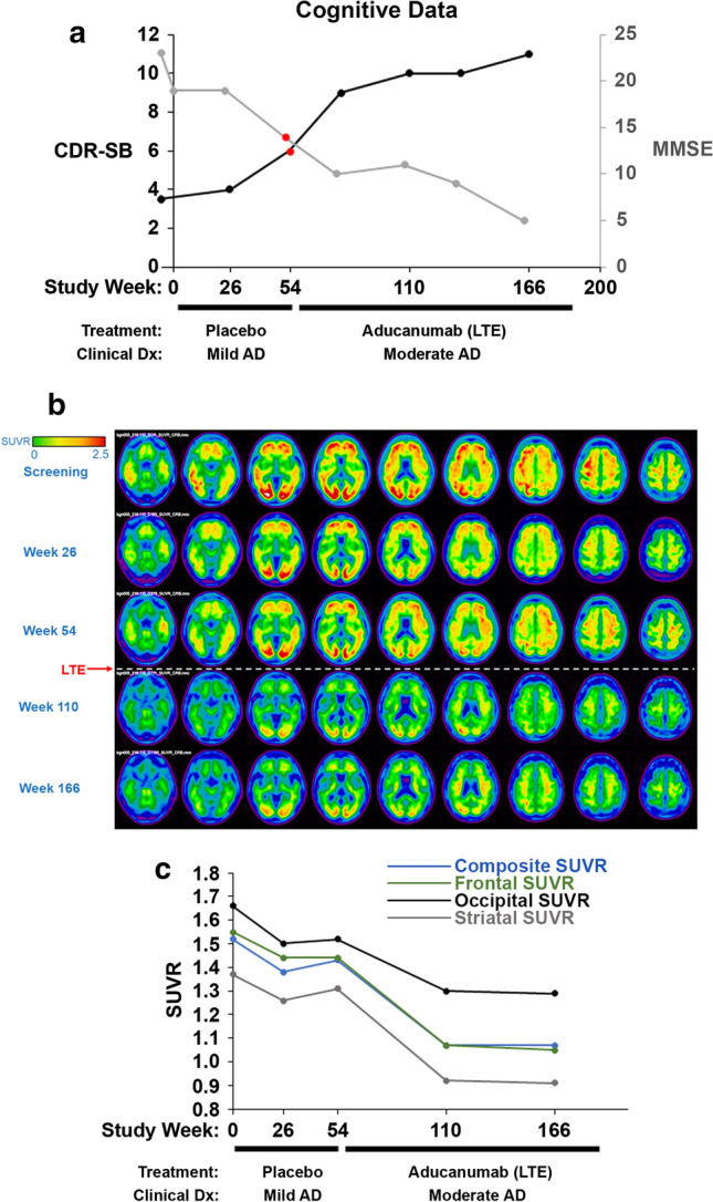
Amyloid beta (Aβ) plaque is a defining pathologic feature of Alzheimer disease (AD). Aducanumab, a monoclonal IgG1 that selectively binds aggregated species of Aβ, has been shown by amyloid positron emission tomography (Amyloid PET) to reduce Aβ plaques in patients with prodromal and mild AD. This is the first autopsy report of the AD neuropathology in a patient previously treated with aducanumab. The patient was an 84-year-old woman who was randomized to the placebo arm of the PRIME Phase 1b study (221AD103). The patient progressed to moderate dementia (MMSE = 14/30), beyond the targeted early AD treatment stage, before receiving aducanumab in the long-term extension (LTE). The patient then received 32 monthly doses of aducanumab, titrated up to 6 mg/kg, for a cumulative dose of 186 mg/kg. In the LTE, Amyloid PET scans demonstrated robust Aβ plaque reduction, from a composite standard uptake value ratio (SUVR) of 1.5 at screening to < 1.1 at 56 weeks post-aducanumab dosing. MRI examinations were negative for amyloid-related imaging abnormalities (ARIA). She passed away in hospice care 4 months after her last dose of aducanumab. The postmortem neuropathologic examination confirmed AD neuropathologic changes. Aβ and IBA1 immunohistochemistry assays demonstrated sparse residual Aβ plaque engaged by amoeboid reactive microglia. Phospho-Tau (pTau) immunohistochemistry demonstrated neocortical neurofibrillary degeneration (Braak stage V, NIA/AA Stage B3). However, the density of pTau neuropathology, including neuritic plaque pTau (NP-Tau), appeared lower in the PRIME LTE Patient compared to a reference cohort of untreated Braak stage V-VI, NIA/AA Stage B3 AD cases. Taken together, this case report is the first to provide Amyloid PET and neuropathologic evidence substantiating the impact of aducanumab to reduce Aβ plaque neuropathology in a patient with AD. Furthermore, this report underscores the critical importance of autopsy neuropathology studies to augment our understanding of aducanumab's mechanism of action and impact on AD biomarkers.
淀粉样蛋白β (Aβ) 斑块是阿尔茨海默病 (AD) 的一种明确的病理特征。Aducanumab 是一种单克隆 IgG1,它选择性地结合 Aβ 的聚集物,通过淀粉样蛋白正电子发射断层扫描 (Amyloid PET) 已被证明可减少前驱期和轻度 AD 患者的 Aβ 斑块。这是首例接受 aducanumab 治疗的 AD 神经病理学的尸检报告。患者是一名 84 岁女性,在 PRIME 1b 期研究 (221AD103) 中被随机分配到安慰剂组。在接受 aducanumab 的长期扩展 (LTE) 之前,患者进展为中度痴呆 (MMSE = 14/30),超出了早期 AD 治疗阶段的目标。然后,患者接受了 32 个月的 aducanumab 剂量,滴定至 6mg/kg,累积剂量为 186mg/kg。在 LTE 中,Amyloid PET 扫描显示 Aβ 斑块有明显减少,从筛查时的复合标准摄取值比值 (SUVR)1.5 降至 aducanumab 给药后 56 周时的 <1.1。MRI 检查未发现与淀粉样蛋白相关的成像异常 (ARIA)。她在接受最后一剂 aducanumab 后 4 个月在临终关怀中心去世。尸检神经病理学检查证实 AD 神经病理学改变。Aβ 和 IBA1 免疫组织化学检测显示稀疏的残留 Aβ 斑块被阿米巴样反应性小胶质细胞所占据。磷酸化 Tau (pTau) 免疫组织化学显示新皮质神经纤维缠结变性 (Braak 阶段 V,NIA/AA 阶段 B3)。然而,与未经治疗的 Braak 阶段 V-VI、NIA/AA 阶段 B3 AD 病例的参考队列相比,PRIME LTE 患者的 pTau 神经病理学密度(包括神经原纤维缠结 pTau (NP-Tau))似乎较低。综上所述,本病例报告首次提供了 Amyloid PET 和神经病理学证据,证实了 aducanumab 可降低 AD 患者的 Aβ 斑块神经病理学。此外,本报告强调了尸检神经病理学研究的重要性,以增加我们对 aducanumab 作用机制和对 AD 生物标志物影响的理解。