Department of Neurosurgery, University of Oklahoma Health Sciences Center, Oklahoma City, Oklahoma, USA.
Omniscient Neurotechnology, Sydney, New South Wales, Australia.
Brain Behav. 2022 Jul;12(7):e2646. doi: 10.1002/brb3.2646. Epub 2022 Jun 22.
The salience network (SN) is a transitory mediator between active and passive states of mind. Multiple cortical areas, including the opercular, insular, and cingulate cortices have been linked in this processing, though knowledge of network connectivity has been devoid of structural specificity.
The current study sought to create an anatomically specific connectivity model of the neural substrates involved in the salience network.
A literature search of PubMed and BrainMap Sleuth was conducted for resting-state and task-based fMRI studies relevant to the salience network according to PRISMA guidelines. Publicly available meta-analytic software was utilized to extract relevant fMRI data for the creation of an activation likelihood estimation (ALE) map and relevant parcellations from the human connectome project overlapping with the ALE data were identified for inclusion in our SN model. DSI-based fiber tractography was then performed on publicaly available data from healthy subjects to determine the structural connections between cortical parcellations comprising the network.
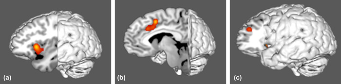
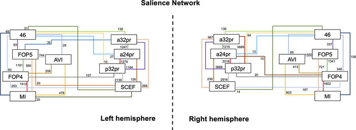
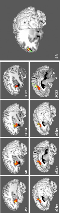
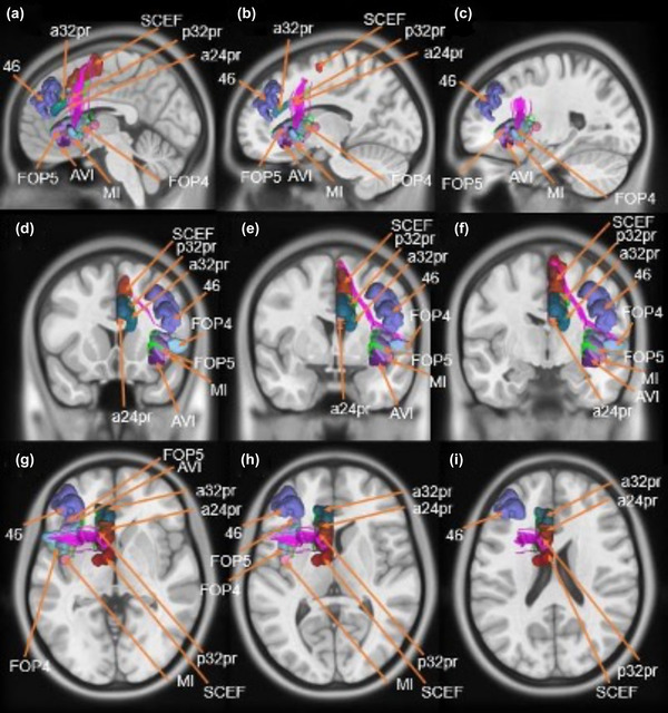
Nine cortical regions were found to comprise the salience network: areas AVI (anterior ventral insula), MI (middle insula), FOP4 (frontal operculum 4), FOP5 (frontal operculum 5), a24pr (anterior 24 prime), a32pr (anterior 32 prime), p32pr (posterior 32 prime), and SCEF (supplementary and cingulate eye field), and 46. The frontal aslant tract was found to connect the opercular-insular cluster to the middle cingulate clusters of the network, while mostly short U-fibers connected adjacent nodes of the network.
Here we provide an anatomically specific connectivity model of the neural substrates involved in the salience network. These results may serve as an empiric basis for clinical translation in this region and for future study which seeks to expand our understanding of how specific neural substrates are involved in salience processing and guide subsequent human behavior.
突显网络(SN)是主动和被动思维状态之间的瞬态中介。多个皮质区域,包括脑岛、岛盖和扣带回皮质,都参与了这一处理过程,但对网络连接的了解缺乏结构特异性。
本研究旨在创建一个涉及突显网络的神经基质的解剖特异性连接模型。
根据 PRISMA 指南,对 PubMed 和 BrainMap Sleuth 进行文献检索,以获取与突显网络相关的静息状态和任务态 fMRI 研究。利用公共可用的元分析软件提取与突显网络相关的 fMRI 数据,以创建激活似然估计(ALE)图,并从人类连接组计划中识别与 ALE 数据重叠的相关分割,以纳入我们的 SN 模型。然后,对来自健康受试者的公共弥散张量成像(DTI)数据进行纤维束追踪,以确定组成网络的皮质分割之间的结构连接。
发现九个皮质区域构成了突显网络:区域 AVI(前腹侧脑岛)、MI(中脑岛)、FOP4(额盖 4)、FOP5(额盖 5)、a24pr(前 24 个)、a32pr(前 32 个)、p32pr(后 32 个)和 SCEF(补充和扣带眼区),以及 46。发现额斜束将脑岛-脑盖簇连接到网络的中扣带回簇,而大多数短 U-纤维连接网络的相邻节点。
在这里,我们提供了一个涉及突显网络的神经基质的解剖特异性连接模型。这些结果可能为该区域的临床转化以及未来旨在扩大我们对特定神经基质如何参与突显处理并指导随后人类行为的理解的研究提供经验基础。