• 文献检索
  • 文档翻译
  • 深度研究
  • 学术资讯
  • Suppr Zotero 插件Zotero 插件
  • 邀请有礼
  • 套餐&价格
  • 历史记录
应用&插件
Suppr Zotero 插件Zotero 插件浏览器插件Mac 客户端Windows 客户端微信小程序
定价
高级版会员购买积分包购买API积分包
服务
文献检索文档翻译深度研究API 文档MCP 服务
关于我们
关于 Suppr公司介绍联系我们用户协议隐私条款
关注我们

Suppr 超能文献

核心技术专利:CN118964589B侵权必究
粤ICP备2023148730 号-1Suppr @ 2026

文献检索

告别复杂PubMed语法,用中文像聊天一样搜索,搜遍4000万医学文献。AI智能推荐,让科研检索更轻松。

立即免费搜索

文件翻译

保留排版,准确专业,支持PDF/Word/PPT等文件格式,支持 12+语言互译。

免费翻译文档

深度研究

AI帮你快速写综述,25分钟生成高质量综述,智能提取关键信息,辅助科研写作。

立即免费体验

新型3D打印甲基丙烯酸化壳聚糖-锂皂石纳米硅酸盐复合支架促进MC3T3前成骨细胞的细胞生长和生物矿化形成。

Novel 3D-printed methacrylated chitosan-laponite nanosilicate composite scaffolds enhance cell growth and biomineral formation in MC3T3 pre-osteoblasts.

作者信息

Cebe Tugba, Ahuja Neelam, Monte Felipe, Awad Kamal, Vyavhare Kimaya, Aswath Pranesh, Huang Jian, Brotto Marco, Varanasi Venu

机构信息

Department of Materials Science and Engineering, University of Texas at Arlington, Arlington, Texas 76019, USA.

Department of Graduate Nursing, College of Nursing and Health Innovation, University of Texas at Arlington, Arlington, Texas 76019, USA.

出版信息

J Mater Res. 2020 Jan;35(1):58-75. doi: 10.1557/jmr.2018.260. Epub 2020 Jan 1.

DOI:10.1557/jmr.2018.260
PMID:35844898
原文链接:https://pmc.ncbi.nlm.nih.gov/articles/PMC9285673/
Abstract

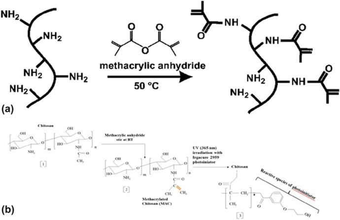
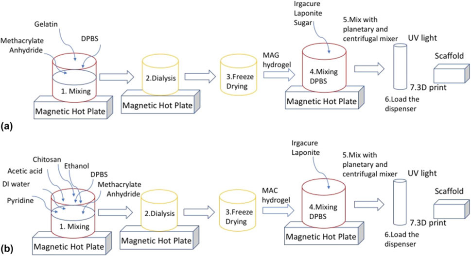
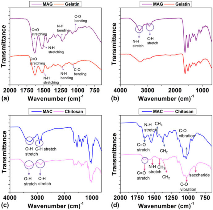
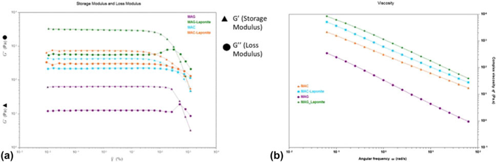
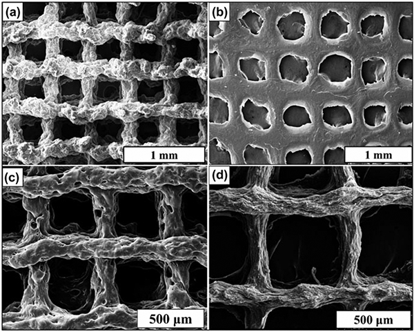
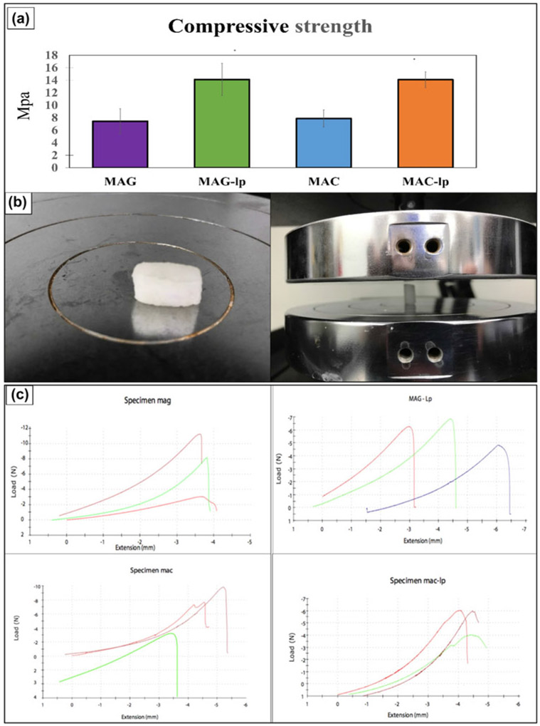
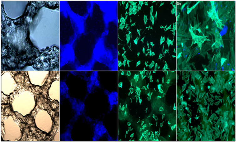
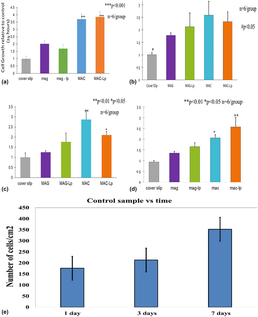
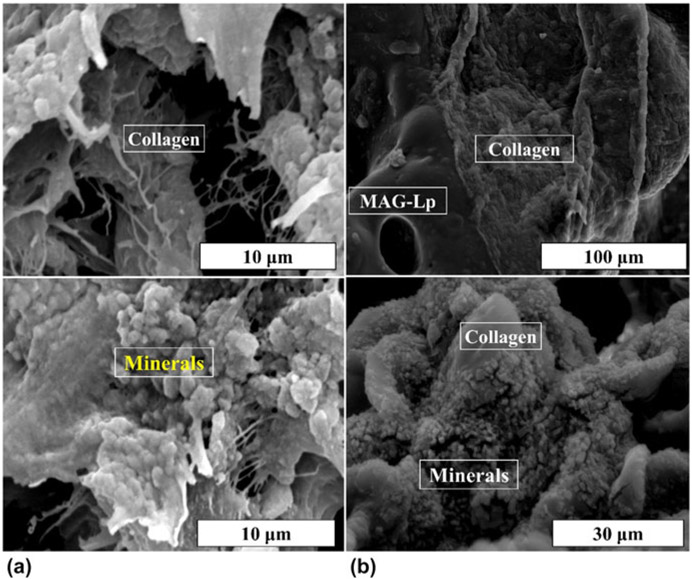
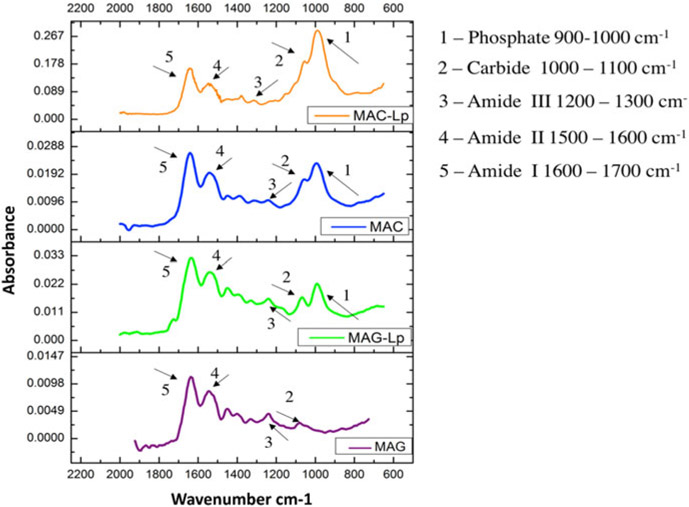
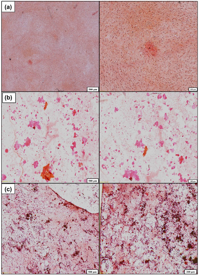
This study compared the effect of gelatin- and chitosan-based scaffolds on osteoblast biomineralization. These scaffolds have been modified using methacrylate and laponite nanosilicates to improve their mechanical strength and support osteoblast function. Scaffold materials were prepared to have the same compressive strength (14-15 MPa) such that differences in cell response would be isolated to differences in biopolymer chemistry. The materials were tested for rheological properties to optimize the bio-ink for successful 3D printing using a robocast-assisted deposition system. Osteoblasts were cultured on the surface of 3D-printed methacrylated chitosan-laponite (MAC-Lp), methacrylated gelatin-laponite (MAG-Lp), MAC, and MAG scaffolds. MAC-Lp scaffolds showed increased cell viability, cell growth, and biomineral formation as compared to MAG-Lp scaffolds. FTIR results showed the presence of higher biomineral phosphate and extracellular matrix (ECM) collagen-like amide formation on MAC-Lp scaffolds as compared to MAG-Lp scaffolds. MAC-Lp scaffolds showed increased density of ECM-like tissue from SEM analysis, stained mineral nodules from Alizarin staining, and the existence of Ca─P species evident by X-ray absorbance near edge structure analysis. In conclusion, MAC-Lp scaffolds enhanced osteoblast growth and biomineral formation as compared to MAG-Lp scaffolds.

摘要

本研究比较了明胶基和壳聚糖基支架对成骨细胞生物矿化的影响。这些支架已用甲基丙烯酸酯和锂皂石纳米硅酸盐进行了改性,以提高其机械强度并支持成骨细胞功能。制备的支架材料具有相同的抗压强度(14 - 15兆帕),以便将细胞反应的差异分离为生物聚合物化学组成的差异。对这些材料进行流变学性能测试,以优化生物墨水,以便使用机器人铸造辅助沉积系统成功进行3D打印。将成骨细胞培养在3D打印的甲基丙烯酸化壳聚糖 - 锂皂石(MAC - Lp)、甲基丙烯酸化明胶 - 锂皂石(MAG - Lp)、MAC和MAG支架表面。与MAG - Lp支架相比,MAC - Lp支架显示出更高的细胞活力、细胞生长和生物矿化形成。傅里叶变换红外光谱(FTIR)结果表明,与MAG - Lp支架相比,MAC - Lp支架上存在更高的生物矿化磷酸盐和细胞外基质(ECM)胶原样酰胺形成。扫描电子显微镜(SEM)分析显示MAC - Lp支架上类似ECM的组织密度增加,茜素红染色显示有染色的矿化结节,X射线吸收近边结构分析表明存在钙磷物种。总之,与MAG - Lp支架相比,MAC - Lp支架增强了成骨细胞生长和生物矿化形成。

https://cdn.ncbi.nlm.nih.gov/pmc/blobs/2f12/9285673/0bf8dcb18be7/nihms-981011-f0012.jpg
https://cdn.ncbi.nlm.nih.gov/pmc/blobs/2f12/9285673/c39eadec264f/nihms-981011-f0001.jpg
https://cdn.ncbi.nlm.nih.gov/pmc/blobs/2f12/9285673/1274c2bcc80b/nihms-981011-f0002.jpg
https://cdn.ncbi.nlm.nih.gov/pmc/blobs/2f12/9285673/7f682e1f2bc2/nihms-981011-f0003.jpg
https://cdn.ncbi.nlm.nih.gov/pmc/blobs/2f12/9285673/785bb59690dc/nihms-981011-f0004.jpg
https://cdn.ncbi.nlm.nih.gov/pmc/blobs/2f12/9285673/d614b83dc69a/nihms-981011-f0005.jpg
https://cdn.ncbi.nlm.nih.gov/pmc/blobs/2f12/9285673/761b0250b25e/nihms-981011-f0006.jpg
https://cdn.ncbi.nlm.nih.gov/pmc/blobs/2f12/9285673/14c76e4b24c4/nihms-981011-f0007.jpg
https://cdn.ncbi.nlm.nih.gov/pmc/blobs/2f12/9285673/d9ca4313355e/nihms-981011-f0008.jpg
https://cdn.ncbi.nlm.nih.gov/pmc/blobs/2f12/9285673/511e2661ef27/nihms-981011-f0009.jpg
https://cdn.ncbi.nlm.nih.gov/pmc/blobs/2f12/9285673/8ff5bbec135d/nihms-981011-f0010.jpg
https://cdn.ncbi.nlm.nih.gov/pmc/blobs/2f12/9285673/5f129881dcfc/nihms-981011-f0011.jpg
https://cdn.ncbi.nlm.nih.gov/pmc/blobs/2f12/9285673/0bf8dcb18be7/nihms-981011-f0012.jpg

相似文献

1
Novel 3D-printed methacrylated chitosan-laponite nanosilicate composite scaffolds enhance cell growth and biomineral formation in MC3T3 pre-osteoblasts.新型3D打印甲基丙烯酸化壳聚糖-锂皂石纳米硅酸盐复合支架促进MC3T3前成骨细胞的细胞生长和生物矿化形成。
J Mater Res. 2020 Jan;35(1):58-75. doi: 10.1557/jmr.2018.260. Epub 2020 Jan 1.
2
Preparation, characterization and bioactivities of nano anhydrous calcium phosphate added gelatin-chitosan scaffolds for bone tissue engineering.纳米无水磷酸钙添加明胶-壳聚糖支架的制备、表征及生物活性研究及其在骨组织工程中的应用。
J Biomater Sci Polym Ed. 2019 Dec;30(18):1756-1778. doi: 10.1080/09205063.2019.1663474. Epub 2019 Sep 17.
3
A natural biomineral for enhancing the biomineralization and cell response of 3D printed polylactic acid bone scaffolds.一种天然生物矿物,用于增强 3D 打印聚乳酸骨支架的生物矿化和细胞反应。
Int J Biol Macromol. 2023 Jul 1;242(Pt 1):124728. doi: 10.1016/j.ijbiomac.2023.124728. Epub 2023 May 5.
4
3D Printed Chitosan Composite Scaffold for Chondrocytes Differentiation.3D 打印壳聚糖复合支架促进软骨细胞分化。
Curr Med Imaging. 2021;17(7):832-842. doi: 10.2174/1573405616666201217112939.
5
Enhanced osteogenic activity by MC3T3-E1 pre-osteoblasts on chemically surface-modified poly(ε-caprolactone) 3D-printed scaffolds compared to RGD immobilized scaffolds.MC3T3-E1 前成骨细胞在化学表面改性聚己内酯 3D 打印支架上的增强成骨活性优于 RGD 固定支架。
Biomed Mater. 2018 Nov 13;14(1):015008. doi: 10.1088/1748-605X/aaeb82.
6
Control of maleic acid-propylene diepoxide hydrogel for 3D printing application for flexible tissue engineering scaffold with high resolution by end capping and graft polymerization.通过封端和接枝聚合控制马来酸-环氧丙烷二环氧物水凝胶用于3D打印应用,以制备具有高分辨率的柔性组织工程支架。
Biomater Res. 2022 Dec 9;26(1):75. doi: 10.1186/s40824-022-00318-x.
7
Sulfated carboxymethyl cellulose and carboxymethyl κ-carrageenan immobilization on 3D-printed poly-ε-caprolactone scaffolds differentially promote pre-osteoblast proliferation and osteogenic activity.硫酸化羧甲基纤维素和羧甲基κ-卡拉胶固定在3D打印聚ε-己内酯支架上对前成骨细胞增殖和成骨活性的促进作用存在差异。
Front Bioeng Biotechnol. 2022 Sep 23;10:957263. doi: 10.3389/fbioe.2022.957263. eCollection 2022.
8
Investigating the mechanical, physiochemical and osteogenic properties in gelatin-chitosan-bioactive nanoceramic composite scaffolds for bone tissue regeneration: In vitro and in vivo.研究明胶-壳聚糖-生物活性纳米陶瓷复合支架的力学、物理化学和成骨性能,用于骨组织再生:体外和体内。
Mater Sci Eng C Mater Biol Appl. 2019 Jan 1;94:713-728. doi: 10.1016/j.msec.2018.10.022. Epub 2018 Oct 4.
9
Proliferation and differentiation of mesenchymal stem cells on scaffolds containing chitosan, calcium polyphosphate and pigeonite for bone tissue engineering.间充质干细胞在含壳聚糖、聚磷酸钙和透辉石的骨组织工程支架上的增殖与分化
Cell Prolif. 2018 Feb;51(1). doi: 10.1111/cpr.12408. Epub 2017 Nov 21.
10
[Osteogenesis effect of dynamic mechanical loading on MC3T3-E1 cells in three-dimensional printing biomimetic composite scaffolds].动态机械加载对三维打印仿生复合支架中MC3T3-E1细胞的成骨作用
Zhongguo Xiu Fu Chong Jian Wai Ke Za Zhi. 2018 Apr 15;32(4):448-456. doi: 10.7507/1002-1892.201711091.

引用本文的文献

1
Extracellular-Matrix-Mimetic Hydrogels by Using Nanomaterials.利用纳米材料制备的细胞外基质模拟水凝胶
Int J Mol Sci. 2025 May 22;26(11):4987. doi: 10.3390/ijms26114987.
2
Three-Dimensional Printing of Hydrogel Blend Tissue Engineering Scaffolds with In Situ Delivery of Anticancer Drug for Treating Melanoma Resection-Induced Tissue Defects.用于治疗黑色素瘤切除诱导组织缺损的原位递送抗癌药物的水凝胶混合组织工程支架的三维打印
J Funct Biomater. 2024 Dec 18;15(12):381. doi: 10.3390/jfb15120381.
3
Nanomaterials-incorporated hydrogels for 3D bioprinting technology.

本文引用的文献

1
Bioactive nanoengineered hydrogels for bone tissue engineering: a growth-factor-free approach.用于骨组织工程的生物活性纳米工程水凝胶:一种无生长因子的方法。
ACS Nano. 2015 Mar 24;9(3):3109-18. doi: 10.1021/nn507488s. Epub 2015 Feb 25.
2
Bone apatite composition of necrotic trabecular bone in the femoral head of immature piglets.未成熟仔猪股骨头坏死小梁骨的骨磷灰石成分
Calcif Tissue Int. 2015 Apr;96(4):324-34. doi: 10.1007/s00223-015-9959-7. Epub 2015 Feb 7.
3
Porosity and dielectric properties as tools to predict drug release trends from hydrogels.
用于3D生物打印技术的纳米材料复合水凝胶
Nano Converg. 2023 Nov 15;10(1):52. doi: 10.1186/s40580-023-00402-5.
4
Biomimetic chitosan with biocomposite nanomaterials for bone tissue repair and regeneration.用于骨组织修复与再生的具有生物复合纳米材料的仿生壳聚糖。
Beilstein J Nanotechnol. 2022 Sep 29;13:1051-1067. doi: 10.3762/bjnano.13.92. eCollection 2022.
5
Two-dimensional nanomaterials-added dynamism in 3D printing and bioprinting of biomedical platforms: Unique opportunities and challenges.二维纳米材料在 3D 打印和生物打印生物医学平台中的动态作用:独特的机遇和挑战。
Biomaterials. 2022 May;284:121507. doi: 10.1016/j.biomaterials.2022.121507. Epub 2022 Apr 5.
6
Interfacial adhesion and surface bioactivity of anodized titanium modified with SiON and SiONP surface coatings.用SiON和SiONP表面涂层改性的阳极氧化钛的界面附着力和表面生物活性。
Surf Interfaces. 2022 Feb;28. doi: 10.1016/j.surfin.2021.101645. Epub 2021 Nov 28.
7
Hypoxia-mimicking 3D bioglass-nanoclay scaffolds promote endogenous bone regeneration.缺氧模拟三维生物玻璃-纳米粘土支架促进内源性骨再生。
Bioact Mater. 2021 Mar 21;6(10):3485-3495. doi: 10.1016/j.bioactmat.2021.03.011. eCollection 2021 Oct.
8
Ionic Silicon Protects Oxidative Damage and Promotes Skeletal Muscle Cell Regeneration.离子硅保护氧化损伤,促进骨骼肌细胞再生。
Int J Mol Sci. 2021 Jan 6;22(2):497. doi: 10.3390/ijms22020497.
9
Inspired by nature: facile design of nanoclay-organic hydrogel bone sealant with multifunctional properties for robust bone regeneration.受自然启发:用于强健骨再生的具有多功能特性的纳米粘土-有机水凝胶骨密封剂的简易设计
Adv Funct Mater. 2020 Oct 22;30(43). doi: 10.1002/adfm.202003717. Epub 2020 Sep 2.
10
Three-Dimensional Printing Constructs Based on the Chitosan for Tissue Regeneration: State of the Art, Developing Directions and Prospect Trends.基于壳聚糖的三维打印构建体用于组织再生:现状、发展方向与前景趋势
Materials (Basel). 2020 Jun 11;13(11):2663. doi: 10.3390/ma13112663.
孔隙率和介电性能作为预测水凝胶药物释放趋势的工具。
Springerplus. 2014 Jul 29;3:393. doi: 10.1186/2193-1801-3-393. eCollection 2014.
4
High-water-content mouldable polyvinyl alcohol-borax hydrogels reinforced by well-dispersed cellulose nanoparticles: dynamic rheological properties and hydrogel formation mechanism.高含水量可模塑的聚乙烯醇-硼砂水凝胶,由分散良好的纤维素纳米颗粒增强:动态流变性能和水凝胶形成机理。
Carbohydr Polym. 2014 Feb 15;102:306-16. doi: 10.1016/j.carbpol.2013.11.045. Epub 2013 Dec 4.
5
Rheology and adhesion of poly(acrylic acid)/laponite nanocomposite hydrogels as biocompatible adhesives.聚(丙烯酸)/锂蒙脱石纳米复合水凝胶的流变学和粘附性作为生物相容性粘合剂。
Langmuir. 2014 Feb 18;30(6):1636-42. doi: 10.1021/la4045623. Epub 2014 Feb 3.
6
Autograft, allograft and bone substitutes in reconstructive orthopedic surgery.自体骨、同种异体骨和骨替代物在矫形骨科手术中的应用。
Aging Clin Exp Res. 2013 Oct;25 Suppl 1:S101-3. doi: 10.1007/s40520-013-0088-8. Epub 2013 Sep 18.
7
XANES analysis of dried and calcined bones.XANES 分析干燥和煅烧的骨骼。
Mater Sci Eng C Mater Biol Appl. 2013 Oct;33(7):3968-79. doi: 10.1016/j.msec.2013.05.038. Epub 2013 May 25.
8
Bioactive silicate nanoplatelets for osteogenic differentiation of human mesenchymal stem cells.用于人骨髓间充质干细胞成骨分化的生物活性硅酸纳米片。
Adv Mater. 2013 Jun 25;25(24):3329-36. doi: 10.1002/adma.201300584. Epub 2013 May 13.
9
Physically crosslinked nanocomposites from silicate-crosslinked PEO: mechanical properties and osteogenic differentiation of human mesenchymal stem cells.基于硅酸盐交联聚氧化乙烯的物理交联纳米复合材料:人骨髓间充质干细胞的机械性能和成骨分化。
Macromol Biosci. 2012 Jun;12(6):779-93. doi: 10.1002/mabi.201100508. Epub 2012 Apr 19.
10
Increased matrix synthesis by fibroblasts with decreased proliferation on synthetic chitosan-gelatin porous structures.成纤维细胞在合成壳聚糖-明胶多孔结构上增殖减少时,基质合成增加。
Biotechnol Bioeng. 2012 May;109(5):1314-25. doi: 10.1002/bit.24396. Epub 2011 Dec 13.