• 文献检索
  • 文档翻译
  • 深度研究
  • 学术资讯
  • Suppr Zotero 插件Zotero 插件
  • 邀请有礼
  • 套餐&价格
  • 历史记录
应用&插件
Suppr Zotero 插件Zotero 插件浏览器插件Mac 客户端Windows 客户端微信小程序
定价
高级版会员购买积分包购买API积分包
服务
文献检索文档翻译深度研究API 文档MCP 服务
关于我们
关于 Suppr公司介绍联系我们用户协议隐私条款
关注我们

Suppr 超能文献

核心技术专利:CN118964589B侵权必究
粤ICP备2023148730 号-1Suppr @ 2026

文献检索

告别复杂PubMed语法,用中文像聊天一样搜索,搜遍4000万医学文献。AI智能推荐,让科研检索更轻松。

立即免费搜索

文件翻译

保留排版,准确专业,支持PDF/Word/PPT等文件格式,支持 12+语言互译。

免费翻译文档

深度研究

AI帮你快速写综述,25分钟生成高质量综述,智能提取关键信息,辅助科研写作。

立即免费体验

利用深度学习技术从急诊初始胸部 X 光片中自动诊断和预测 COVID-19 肺炎。

Automated diagnosis and prognosis of COVID-19 pneumonia from initial ER chest X-rays using deep learning.

机构信息

Department of Radiology and Radiologic Sciences, Division of Cardiothoracic Radiology, Medical University of South Carolina, Charleston, SC, USA.

Siemens Healthineers, Malvern, PA, USA.

出版信息

BMC Infect Dis. 2022 Jul 21;22(1):637. doi: 10.1186/s12879-022-07617-7.

DOI:10.1186/s12879-022-07617-7
PMID:35864468
原文链接:https://pmc.ncbi.nlm.nih.gov/articles/PMC9301895/
Abstract

BACKGROUND

Airspace disease as seen on chest X-rays is an important point in triage for patients initially presenting to the emergency department with suspected COVID-19 infection. The purpose of this study is to evaluate a previously trained interpretable deep learning algorithm for the diagnosis and prognosis of COVID-19 pneumonia from chest X-rays obtained in the ED.

METHODS

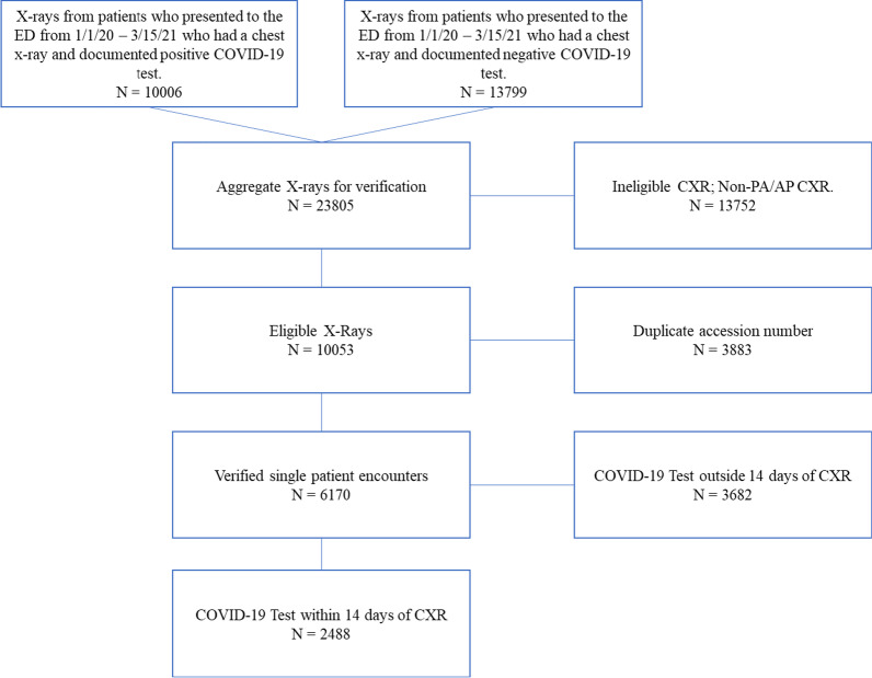
This retrospective study included 2456 (50% RT-PCR positive for COVID-19) adult patients who received both a chest X-ray and SARS-CoV-2 RT-PCR test from January 2020 to March of 2021 in the emergency department at a single U.S.

INSTITUTION

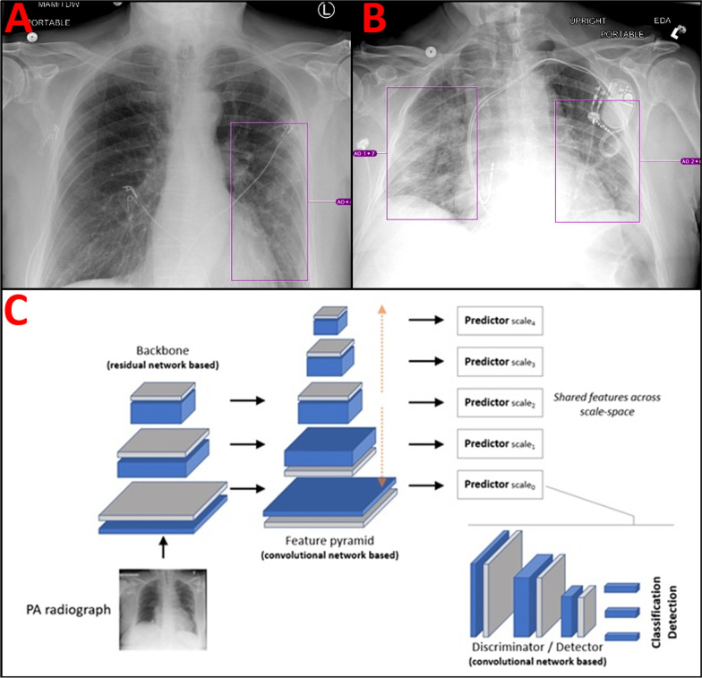
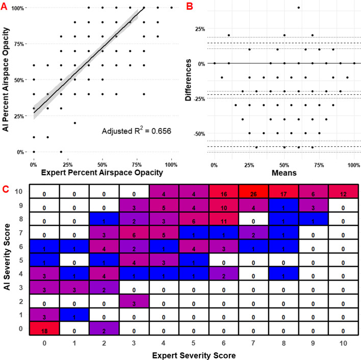
A total of 2000 patients were included as an additional training cohort and 456 patients in the randomized internal holdout testing cohort for a previously trained Siemens AI-Radiology Companion deep learning convolutional neural network algorithm. Three cardiothoracic fellowship-trained radiologists systematically evaluated each chest X-ray and generated an airspace disease area-based severity score which was compared against the same score produced by artificial intelligence. The interobserver agreement, diagnostic accuracy, and predictive capability for inpatient outcomes were assessed. Principal statistical tests used in this study include both univariate and multivariate logistic regression.

RESULTS

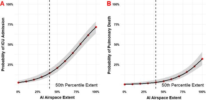
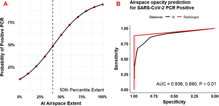
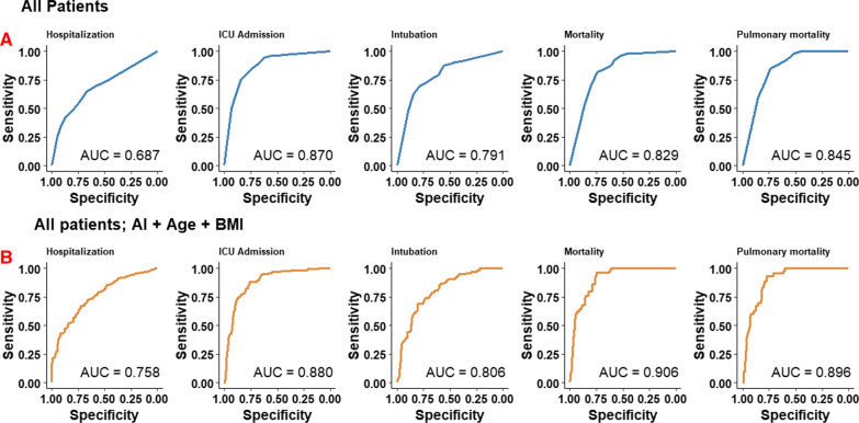
Overall ICC was 0.820 (95% CI 0.790-0.840). The diagnostic AUC for SARS-CoV-2 RT-PCR positivity was 0.890 (95% CI 0.861-0.920) for the neural network and 0.936 (95% CI 0.918-0.960) for radiologists. Airspace opacities score by AI alone predicted ICU admission (AUC = 0.870) and mortality (0.829) in all patients. Addition of age and BMI into a multivariate log model improved mortality prediction (AUC = 0.906).

CONCLUSION

The deep learning algorithm provides an accurate and interpretable assessment of the disease burden in COVID-19 pneumonia on chest radiographs. The reported severity scores correlate with expert assessment and accurately predicts important clinical outcomes. The algorithm contributes additional prognostic information not currently incorporated into patient management.

摘要

背景

在胸部 X 光片上观察到的气腔疾病是对最初因疑似 COVID-19 感染而到急诊科就诊的患者进行分诊的一个重要要点。本研究的目的是评估之前经过训练的可解释深度学习算法,用于诊断和预测急诊科获得的 COVID-19 肺炎的胸部 X 光片。

方法

本回顾性研究纳入了 2020 年 1 月至 2021 年 3 月期间在美国一家医院急诊科接受胸部 X 光检查和 SARS-CoV-2 RT-PCR 检测的 2456 名(50% RT-PCR 检测 COVID-19 阳性)成年患者。共纳入 2000 名患者作为额外的训练队列,456 名患者在随机内部保留测试队列中接受了之前经过训练的西门子 AI-Radiology Companion 深度学习卷积神经网络算法的测试。三名心胸科研究员对每一张胸部 X 光片进行了系统评估,并生成了基于气腔疾病面积的严重程度评分,该评分与人工智能生成的评分进行了比较。评估了观察者间的一致性、诊断准确性和住院患者结局的预测能力。本研究中使用的主要统计检验包括单变量和多变量逻辑回归。

结果

总体 ICC 为 0.820(95% CI 0.790-0.840)。对于 SARS-CoV-2 RT-PCR 阳性,神经网络的诊断 AUC 为 0.890(95% CI 0.861-0.920),放射科医生的 AUC 为 0.936(95% CI 0.918-0.960)。仅使用 AI 进行的气腔混浊评分可预测所有患者的 ICU 入院(AUC=0.870)和死亡率(0.829)。将年龄和 BMI 纳入多变量 log 模型可改善死亡率预测(AUC=0.906)。

结论

深度学习算法可对胸部 X 光片上 COVID-19 肺炎的疾病负担进行准确且可解释的评估。报告的严重程度评分与专家评估相关,可准确预测重要的临床结局。该算法提供了当前患者管理中未包含的额外预后信息。

https://cdn.ncbi.nlm.nih.gov/pmc/blobs/41ca/9306047/f581161bebc5/12879_2022_7617_Fig6_HTML.jpg
https://cdn.ncbi.nlm.nih.gov/pmc/blobs/41ca/9306047/14811ba5bded/12879_2022_7617_Fig1_HTML.jpg
https://cdn.ncbi.nlm.nih.gov/pmc/blobs/41ca/9306047/1c3b85676f13/12879_2022_7617_Fig2_HTML.jpg
https://cdn.ncbi.nlm.nih.gov/pmc/blobs/41ca/9306047/dd1788d96132/12879_2022_7617_Fig3_HTML.jpg
https://cdn.ncbi.nlm.nih.gov/pmc/blobs/41ca/9306047/1f495271dc36/12879_2022_7617_Fig4_HTML.jpg
https://cdn.ncbi.nlm.nih.gov/pmc/blobs/41ca/9306047/ef7ca2778132/12879_2022_7617_Fig5_HTML.jpg
https://cdn.ncbi.nlm.nih.gov/pmc/blobs/41ca/9306047/f581161bebc5/12879_2022_7617_Fig6_HTML.jpg

相似文献

1
Automated diagnosis and prognosis of COVID-19 pneumonia from initial ER chest X-rays using deep learning.利用深度学习技术从急诊初始胸部 X 光片中自动诊断和预测 COVID-19 肺炎。
BMC Infect Dis. 2022 Jul 21;22(1):637. doi: 10.1186/s12879-022-07617-7.
2
Prognostication of patients with COVID-19 using artificial intelligence based on chest x-rays and clinical data: a retrospective study.基于胸部 X 光片和临床数据的人工智能预测 COVID-19 患者的预后:一项回顾性研究。
Lancet Digit Health. 2021 May;3(5):e286-e294. doi: 10.1016/S2589-7500(21)00039-X. Epub 2021 Mar 24.
3
An Interpretable Chest CT Deep Learning Algorithm for Quantification of COVID-19 Lung Disease and Prediction of Inpatient Morbidity and Mortality.一种用于 COVID-19 肺部疾病定量和预测住院患者发病率及死亡率的可解释胸部 CT 深度学习算法。
Acad Radiol. 2022 Aug;29(8):1178-1188. doi: 10.1016/j.acra.2022.03.023. Epub 2022 Apr 4.
4
Initial chest radiographs and artificial intelligence (AI) predict clinical outcomes in COVID-19 patients: analysis of 697 Italian patients.初始胸部 X 光片和人工智能 (AI) 预测 COVID-19 患者的临床结局:对 697 例意大利患者的分析。
Eur Radiol. 2021 Mar;31(3):1770-1779. doi: 10.1007/s00330-020-07269-8. Epub 2020 Sep 18.
5
3D CT-Inclusive Deep-Learning Model to Predict Mortality, ICU Admittance, and Intubation in COVID-19 Patients.基于 3D CT 的深度学习模型预测 COVID-19 患者的死亡率、入住 ICU 率和插管率。
J Digit Imaging. 2023 Apr;36(2):603-616. doi: 10.1007/s10278-022-00734-4. Epub 2022 Nov 30.
6
Determination of disease severity in COVID-19 patients using deep learning in chest X-ray images.利用胸部 X 光图像中的深度学习技术确定 COVID-19 患者的疾病严重程度。
Diagn Interv Radiol. 2021 Jan;27(1):20-27. doi: 10.5152/dir.2020.20205.
7
Explainable COVID-19 Detection Based on Chest X-rays Using an End-to-End RegNet Architecture.基于端到端 RegNet 架构的基于胸部 X 光的可解释 COVID-19 检测。
Viruses. 2023 Jun 6;15(6):1327. doi: 10.3390/v15061327.
8
Deep-learning algorithms for the interpretation of chest radiographs to aid in the triage of COVID-19 patients: A multicenter retrospective study.深度学习算法在解读胸部 X 光片中对 COVID-19 患者分诊的辅助作用:一项多中心回顾性研究。
PLoS One. 2020 Nov 24;15(11):e0242759. doi: 10.1371/journal.pone.0242759. eCollection 2020.
9
Artificial Intelligence-assisted chest X-ray assessment scheme for COVID-19.人工智能辅助 COVID-19 胸片评估方案。
Eur Radiol. 2021 Aug;31(8):6039-6048. doi: 10.1007/s00330-020-07628-5. Epub 2021 Jan 20.
10
DeepCOVID-XR: An Artificial Intelligence Algorithm to Detect COVID-19 on Chest Radiographs Trained and Tested on a Large U.S. Clinical Data Set.DeepCOVID-XR:一种人工智能算法,可在美国大型临床数据集上进行训练和测试,用于检测胸部 X 光片上的 COVID-19。
Radiology. 2021 Apr;299(1):E167-E176. doi: 10.1148/radiol.2020203511. Epub 2020 Nov 24.

引用本文的文献

1
A bibliometric analysis of clinical studies on artificial intelligence in emergency medicine.急诊医学中人工智能临床研究的文献计量分析
Medicine (Baltimore). 2025 Jul 11;104(28):e43282. doi: 10.1097/MD.0000000000043282.
2
Metaheuristic optimizers integrated with vision transformer model for severity detection and classification via multimodal COVID-19 images.通过多模态新冠肺炎图像,将元启发式优化器与视觉Transformer模型集成用于严重程度检测和分类。
Sci Rep. 2025 Apr 22;15(1):13941. doi: 10.1038/s41598-025-98593-w.
3
Classification of the ICU Admission for COVID-19 Patients with Transfer Learning Models Using Chest X-Ray Images.

本文引用的文献

1
An Artificial Intelligence-Based Chest X-ray Model on Human Nodule Detection Accuracy From a Multicenter Study.基于人工智能的多中心研究中用于检测人类结节的胸部 X 射线模型。
JAMA Netw Open. 2021 Dec 1;4(12):e2141096. doi: 10.1001/jamanetworkopen.2021.41096.
2
Disease Severity Scoring for COVID-19: A Welcome (Semi)Quantitative Role for Chest Radiography.新型冠状病毒肺炎的疾病严重程度评分:胸部X线摄影迎来(半)定量作用
Radiology. 2021 Dec;301(3):E445. doi: 10.1148/radiol.2021219022.
3
Chest Radiograph Scoring Alone or Combined with Other Risk Scores for Predicting Outcomes in COVID-19: A UK Study.
使用胸部X光图像的迁移学习模型对COVID-19患者的重症监护病房入院情况进行分类
Diagnostics (Basel). 2025 Mar 26;15(7):845. doi: 10.3390/diagnostics15070845.
4
Artificial Intelligence-Empowered Radiology-Current Status and Critical Review.人工智能赋能的放射学——现状与批判性综述
Diagnostics (Basel). 2025 Jan 24;15(3):282. doi: 10.3390/diagnostics15030282.
5
Towards Improved XAI-Based Epidemiological Research into the Next Potential Pandemic.迈向基于可解释人工智能的流行病学研究,以应对下一次潜在的大流行。
Life (Basel). 2024 Jun 21;14(7):783. doi: 10.3390/life14070783.
6
Artificial Intelligence in Optimizing the Functioning of Emergency Departments; a Systematic Review of Current Solutions.人工智能在优化急诊科运作中的应用;当前解决方案的系统综述
Arch Acad Emerg Med. 2024 Jan 27;12(1):e22. doi: 10.22037/aaem.v12i1.2110. eCollection 2024.
7
Comparison of the Discrimination Performance of AI Scoring and the Brixia Score in Predicting COVID-19 Severity on Chest X-Ray Imaging: Diagnostic Accuracy Study.人工智能评分与布里夏评分在胸部X线成像预测COVID-19严重程度中的鉴别性能比较:诊断准确性研究
JMIR Form Res. 2024 Mar 7;8:e46817. doi: 10.2196/46817.
8
Human-AI Symbiosis: A Path Forward to Improve Chest Radiography and the Role of Radiologists in Patient Care.人机共生:改善胸部X光检查及放射科医生在患者护理中作用的前进道路。
Radiology. 2024 Jan;310(1):e232778. doi: 10.1148/radiol.232778.
9
Unraveling complex relationships between COVID-19 risk factors using machine learning based models for predicting mortality of hospitalized patients and identification of high-risk group: a large retrospective study.使用基于机器学习的模型来揭示新冠病毒疾病(COVID-19)风险因素之间的复杂关系,以预测住院患者的死亡率并识别高危人群:一项大型回顾性研究
Front Med (Lausanne). 2023 May 4;10:1170331. doi: 10.3389/fmed.2023.1170331. eCollection 2023.
10
Comprehensive analysis of clinical data for COVID-19 outcome estimation with machine learning models.使用机器学习模型对COVID-19结果估计的临床数据进行综合分析。
Biomed Signal Process Control. 2023 Jul;84:104818. doi: 10.1016/j.bspc.2023.104818. Epub 2023 Mar 9.
胸部X光片评分单独或联合其他风险评分预测COVID-19患者的预后:一项英国研究。
Radiology. 2021 Dec;301(3):E444. doi: 10.1148/radiol.2021219021.
4
Artificial intelligence-driven assessment of radiological images for COVID-19.人工智能驱动的 COVID-19 放射影像评估。
Comput Biol Med. 2021 Sep;136:104665. doi: 10.1016/j.compbiomed.2021.104665. Epub 2021 Jul 21.
5
Machine learning application for the prediction of SARS-CoV-2 infection using blood tests and chest radiograph.利用血液检测和胸部 X 光预测 SARS-CoV-2 感染的机器学习应用。
Sci Rep. 2021 Jul 9;11(1):14250. doi: 10.1038/s41598-021-93719-2.
6
Checklist for Artificial Intelligence in Medical Imaging (CLAIM): A Guide for Authors and Reviewers.医学影像人工智能清单(CLAIM):作者和审稿人指南
Radiol Artif Intell. 2020 Mar 25;2(2):e200029. doi: 10.1148/ryai.2020200029. eCollection 2020 Mar.
7
Automated Assessment and Tracking of COVID-19 Pulmonary Disease Severity on Chest Radiographs using Convolutional Siamese Neural Networks.使用卷积暹罗神经网络对胸部X光片上的COVID-19肺部疾病严重程度进行自动评估和跟踪。
Radiol Artif Intell. 2020 Jul 22;2(4):e200079. doi: 10.1148/ryai.2020200079. eCollection 2020 Jul.
8
Determinants of Chest X-Ray Sensitivity for COVID- 19: A Multi-Institutional Study in the United States.COVID-19胸部X光检查敏感性的决定因素:美国的一项多机构研究。
Radiol Cardiothorac Imaging. 2020 Sep 24;2(5):e200337. doi: 10.1148/ryct.2020200337. eCollection 2020 Oct.
9
A Characteristic Chest Radiographic Pattern in the Setting of the COVID-19 Pandemic.COVID-19大流行背景下的一种特征性胸部X线表现模式。
Radiol Cardiothorac Imaging. 2020 Sep 3;2(5):e200280. doi: 10.1148/ryct.2020200280. eCollection 2020 Oct.
10
Chest Imaging Appearance of COVID-19 Infection.新型冠状病毒肺炎感染的胸部影像学表现。
Radiol Cardiothorac Imaging. 2020 Feb 13;2(1):e200028. doi: 10.1148/ryct.2020200028. eCollection 2020 Feb.