Neurochemistry Laboratory, Clinical Chemistry department, Amsterdam Neuroscience, VU University Medical Center, Amsterdam UMC, Amsterdam, The Netherlands.
Department of Pathology, Amsterdam Neuroscience, VU University Medical Center, Amsterdam UMC, Amsterdam, The Netherlands.
Alzheimers Res Ther. 2022 Jul 25;14(1):100. doi: 10.1186/s13195-022-01039-y.
YKL-40 (Chitinase 3-like I) is increased in CSF of Alzheimer's disease (AD) and frontotemporal lobar degeneration (FTLD) patients and is therefore considered a potential neuroinflammatory biomarker. Whether changed YKL-40 levels in the CSF reflect dysregulation of YKL-40 in the brain is not completely understood yet. We aimed to extensively analyze YKL-40 levels in the brain of AD and different FTLD pathological subtypes. The direct relationship between YKL-40 levels in post-mortem brain and ante-mortem CSF was examined in a small set of paired brain-CSF samples.
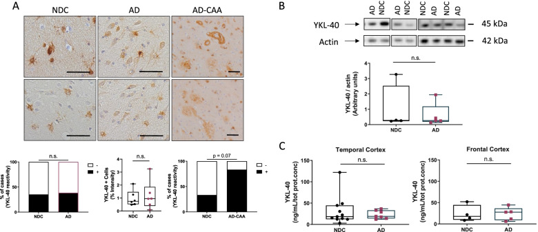
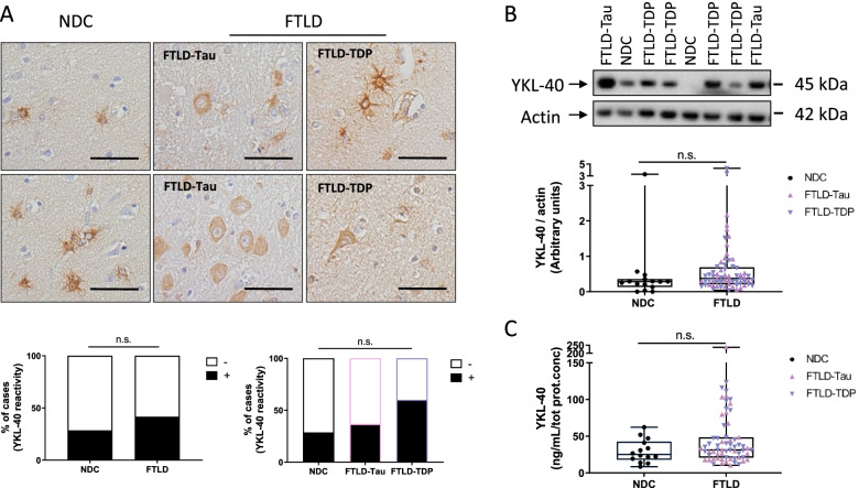
YKL-40 was analyzed in post-mortem temporal and frontal cortex of non-demented controls and patients with AD and FTLD (including FTLD-Tau and FTLD-TDP) pathology by immunohistochemistry (temporal cortex: 51 controls and 56 AD and frontal cortex: 7 controls and 24 FTLD patients), western blot (frontal cortex: 14 controls, 5 AD and 67 FTLD patients), or ELISA (temporal cortex: 11 controls and 7 AD and frontal cortex: 14 controls, 5 AD and 67 FTLD patients). YKL-40 levels were also measured in paired post-mortem brain and ante-mortem CSF samples from dementia patients (n = 9, time-interval collection: 1.4 years) by ELISA.
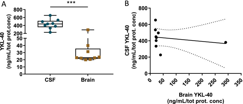
We observed that YKL-40 post-mortem brain levels were similar between AD, FTLD, and controls as shown by immunohistochemistry, western blot, and ELISA. Interestingly, strong YKL-40 immunoreactivity was observed in AD cases with cerebral amyloid angiopathy (CAA; n = 6). In paired CSF-brain samples, YKL-40 concentration was 8-times higher in CSF compared to brain.
Our data suggest that CSF YKL-40 changes may not reflect YKL-40 changes within AD and FTLD pathological brain areas. The YKL-40 reactivity associated with classical CAA hallmarks indicates a possible relationship between YKL-40, neuroinflammation, and vascular pathology.
YKL-40(几丁质酶 3 样蛋白 I)在阿尔茨海默病(AD)和额颞叶变性(FTLD)患者的脑脊液中增加,因此被认为是潜在的神经炎症生物标志物。然而,脑脊液中 YKL-40 水平的变化是否反映了大脑中 YKL-40 的失调尚不完全清楚。我们旨在广泛分析 AD 和不同 FTLD 病理亚型患者大脑中的 YKL-40 水平。我们在一小部分配对的脑-脑脊液样本中检查了死后脑和生前脑脊液中 YKL-40 水平之间的直接关系。
通过免疫组织化学(颞叶皮层:51 名对照和 56 名 AD 和额叶皮层:7 名对照和 24 名 FTLD 患者)、western blot(额叶皮层:14 名对照、5 名 AD 和 67 名 FTLD 患者)或 ELISA(颞叶皮层:11 名对照和 7 名 AD 和额叶皮层:14 名对照、5 名 AD 和 67 名 FTLD 患者)分析非痴呆对照者和 AD 及 FTLD(包括 FTLD-Tau 和 FTLD-TDP)患者死后颞叶和额叶皮质中的 YKL-40。我们还通过 ELISA 测量了痴呆患者死后配对的脑和生前脑脊液样本中的 YKL-40 水平(n=9,时间间隔采集:1.4 年)。
我们观察到,免疫组织化学、western blot 和 ELISA 显示 AD、FTLD 和对照组之间的死后大脑 YKL-40 水平相似。有趣的是,在伴有脑淀粉样血管病(CAA;n=6)的 AD 病例中观察到强烈的 YKL-40 免疫反应。在配对的 CSF-脑样本中,CSF 中的 YKL-40 浓度是脑内的 8 倍。
我们的数据表明,CSF YKL-40 的变化可能无法反映 AD 和 FTLD 病理性脑区的 YKL-40 变化。与经典 CAA 标志物相关的 YKL-40 反应表明 YKL-40、神经炎症和血管病理学之间可能存在关系。