Chunduru Pranathi, Phillips Joanna J, Molinaro Annette M
Department of Neurological Surgery, University of California San Francisco, San Francisco, California, USA.
Neurooncol Adv. 2022 Jul 14;4(1):vdac111. doi: 10.1093/noajnl/vdac111. eCollection 2022 Jan-Dec.
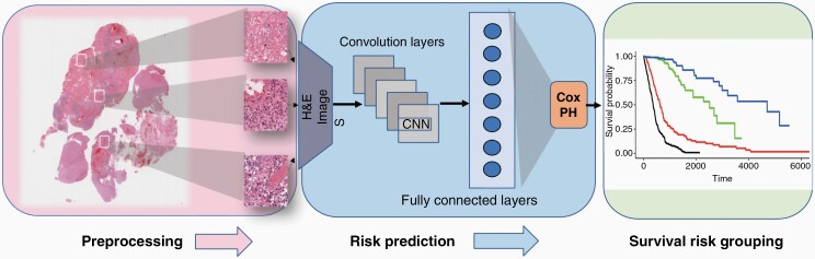
Evaluation of tumor-tissue images stained with hematoxylin and eosin (H&E) is pivotal in diagnosis, yet only a fraction of the rich phenotypic information is considered for clinical care. Here, we propose a survival deep learning (SDL) framework to extract this information to predict glioma survival.
Digitized whole slide images were downloaded from The Cancer Genome Atlas (TCGA) for 766 diffuse glioma patients, including isocitrate dehydrogenase (IDH)-mutant/1p19q-codeleted oligodendroglioma, IDH-mutant/1p19q-intact astrocytoma, and IDH-wildtype astrocytoma/glioblastoma. Our SDL framework employs a residual convolutional neural network with a survival model to predict patient risk from H&E-stained whole-slide images. We used statistical sampling techniques and randomized the transformation of images to address challenges in learning from histology images. The SDL risk score was evaluated in traditional and recursive partitioning (RPA) survival models.
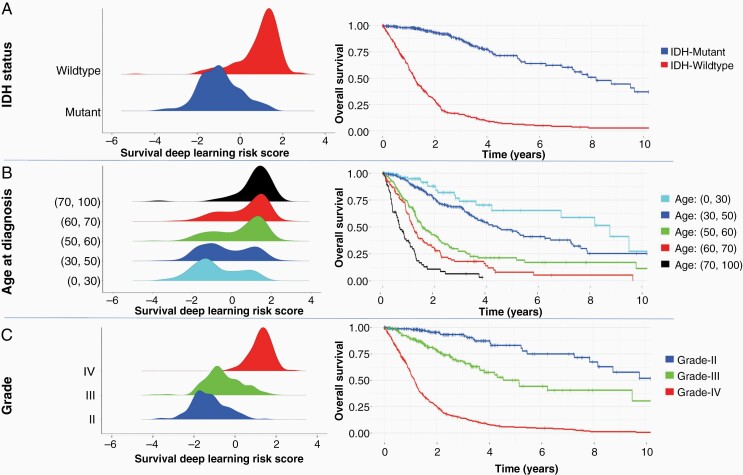
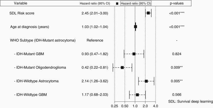
The SDL risk score demonstrated substantial univariate prognostic power (median concordance index of 0.79 [: 0.01]). After adjusting for age and World Health Organization 2016 subtype, the SDL risk score was significantly associated with overall survival (OS; hazard ratio = 2.45; 95% CI: 2.01 to 3.00). Four distinct survival risk groups were characterized by RPA based on SDL risk score, IDH status, and age with markedly different median OS ranging from 1.03 years to 14.14 years.
The present study highlights the independent prognostic power of the SDL risk score for objective and accurate prediction of glioma outcomes. Further, we show that the RPA delineation of patient-specific risk scores and clinical prognostic factors can successfully demarcate the OS of glioma patients.
苏木精和伊红(H&E)染色的肿瘤组织图像评估在诊断中至关重要,但临床诊疗仅考虑了丰富表型信息的一小部分。在此,我们提出一种生存深度学习(SDL)框架来提取这些信息以预测胶质瘤生存情况。
从癌症基因组图谱(TCGA)下载了766例弥漫性胶质瘤患者的数字化全切片图像,包括异柠檬酸脱氢酶(IDH)突变/1p19q共缺失少突胶质细胞瘤、IDH突变/1p19q完整星形细胞瘤以及IDH野生型星形细胞瘤/胶质母细胞瘤。我们的SDL框架采用带有生存模型的残差卷积神经网络,从H&E染色的全切片图像预测患者风险。我们使用统计抽样技术并随机化图像变换,以应对从组织学图像学习中的挑战。在传统和递归划分(RPA)生存模型中评估SDL风险评分。
SDL风险评分显示出显著的单变量预后能力(中位一致性指数为0.79 [: 0.01])。在调整年龄和世界卫生组织2016亚型后,SDL风险评分与总生存期(OS)显著相关(风险比 = 2.45;95%置信区间:2.01至3.00)。基于SDL风险评分、IDH状态和年龄,通过RPA确定了四个不同的生存风险组,中位OS明显不同,范围从1.03年到14.14年。
本研究突出了SDL风险评分在客观准确预测胶质瘤预后方面的独立预后能力。此外,我们表明患者特异性风险评分和临床预后因素的RPA划分能够成功区分胶质瘤患者的OS。