Park Gyusik, Fleifel Mohamad, Kesserwani Hassan N
Neurology, University of Alabama at Birmingham Marnix E. Heersink School of Medicine, Birmingham, USA.
Internal Medicine, American University of Beirut Medical Center, Beirut, LBN.
Cureus. 2022 Aug 1;14(8):e27557. doi: 10.7759/cureus.27557. eCollection 2022 Aug.
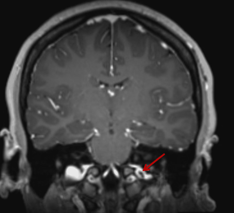
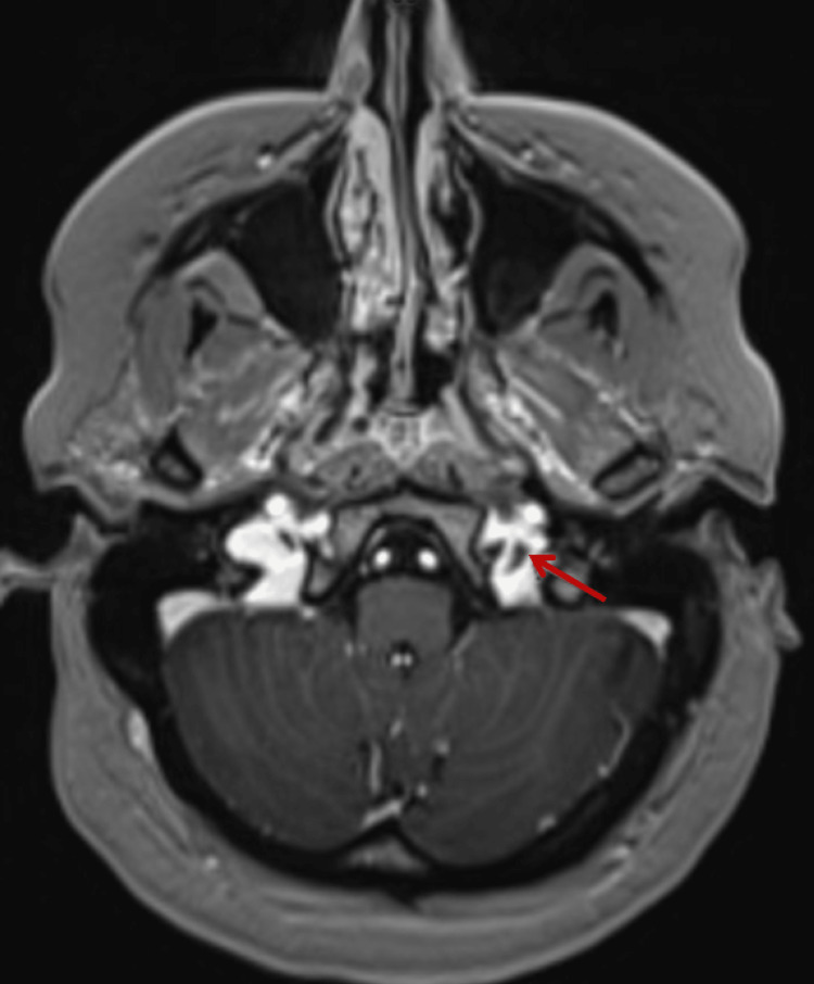
Pseudotumor cerebri (PTC) secondary to cerebral venous sinus thrombosis can be a difficult diagnosis to make for various reasons, including an atypical patient profile and potentially pleomorphic signs and symptoms. The symptoms can be insidious and can evolve acutely, subacutely, or chronically. To complicate the picture even further, neurodiagnostic testing can be particularly troublesome due to both false-positive and false-negative results. Frequently, multiple imaging modalities are variably deployed, and they include computed tomography (CT) with and without contrast, computed tomography venogram (CTV), magnetic resonance imaging (MRI), and magnetic resonance venography (MRV) of the brain. The thrombus can be quite subtle, requiring the seasoned eye of an experienced neuroradiologist. Nevertheless, when a diagnosis is made, the treatment can be highly efficacious and gratifying as it can prevent serious visual complications. We present a rare case of PTC due to a jugular bulb thrombosis and outline the challenging diagnostic steps.
继发于脑静脉窦血栓形成的假性脑瘤(PTC)由于多种原因可能难以诊断,包括不典型的患者情况以及潜在的多样体征和症状。症状可能隐匿,也可能急性、亚急性或慢性进展。更复杂的是,由于假阳性和假阴性结果,神经诊断测试可能特别棘手。通常,会不同程度地采用多种成像方式,包括有或无造影剂的计算机断层扫描(CT)、计算机断层血管造影(CTV)、磁共振成像(MRI)以及脑部磁共振静脉造影(MRV)。血栓可能非常隐匿,需要经验丰富的神经放射科医生的敏锐眼光。然而,一旦做出诊断,治疗可能非常有效且令人满意,因为它可以预防严重的视力并发症。我们报告一例因颈静脉球血栓形成导致的罕见PTC病例,并概述具有挑战性的诊断步骤。