Ding Hao, Yang Yunzhen, Li Xin, Cheung Gary Shun-Pan, Matinlinna Jukka Pekka, Burrow Michael, Tsoi James Kit-Hon
Dental Materials Science, Applied Oral Sciences and Community Dental Care, Faculty of Dentistry, The University of Hong Kong, Pokfulam, Hong Kong.
Restorative Dental Sciences, Faculty of Dentistry, The University of Hong Kong, Pokfulam, Hong Kong.
Biomater Investig Dent. 2022 Aug 31;9(1):75-83. doi: 10.1080/26415275.2022.2114479. eCollection 2022.
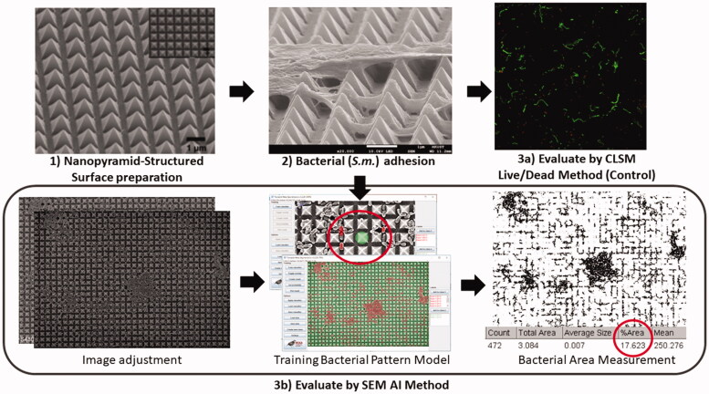
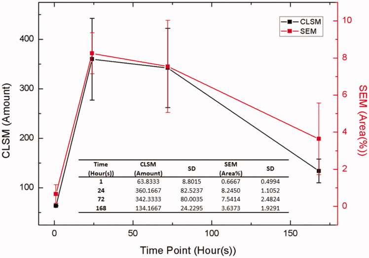
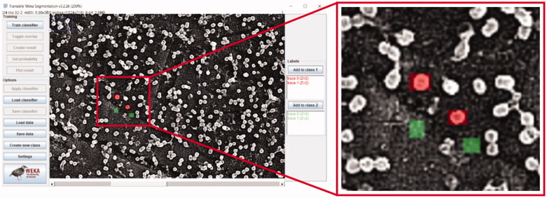
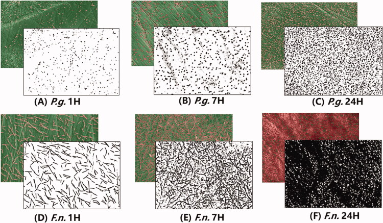
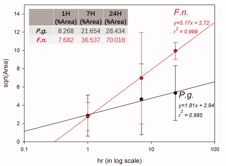
Measurement of bacterial adhesion has been of great interest for different dental materials. Various methods have been used for bacterial counting; however, they are all indirect measurements with estimated results and therefore cannot truly reflect the adhesion status. This study provides a new direct measurement approach by using a simple artificial intelligence (AI) method to quantify the initial bacterial adhesion on different dental materials using Scanning Electron Microscope (SEM) images. () and () were used for bacterial adhesion on dental zirconia surfaces, and the adhesion was evaluated using SEM images at time points of one, seven, and 24 h(s). Image pre-processing and bacterial area measurement were performed using Fiji software with a machine learning plugin. The same AI method was also applied on SEM with () inoculated PMMA nano-structured surface at 1, 24, 72, and 168 h(s), and then further compared with the CLSM method. For both and initiation adhesion on zirconia, a new linear correlation (r > 0.98) was found between bacteria adhered area and time, such that: For S.m. on PMMA surface, live/dead staining CLSM method and the newly proposed AI method on SEM images were strongly and positively associated (Pearson's correlation coefficient > 0.9), i.e. both methods are comparable. SEM images can be analyzed directly for both morphology and quantifying bacterial adhesion on different dental materials' surfaces by the simple AI-enabled method with reduced time, cost, and labours.
细菌黏附的测量一直是不同牙科材料研究的重点。已经使用了各种方法进行细菌计数;然而,它们都是间接测量,结果是估计值,因此不能真实反映黏附状态。本研究提供了一种新的直接测量方法,通过使用一种简单的人工智能(AI)方法,利用扫描电子显微镜(SEM)图像来量化不同牙科材料上的初始细菌黏附情况。()和()用于在牙科氧化锆表面的细菌黏附,并在1、7和24小时的时间点使用SEM图像评估黏附情况。使用带有机器学习插件的Fiji软件进行图像预处理和细菌面积测量。同样的AI方法也应用于接种了()的聚甲基丙烯酸甲酯(PMMA)纳米结构表面的SEM图像,时间点为1、24、72和168小时,然后与共聚焦激光扫描显微镜(CLSM)方法进行进一步比较。对于在氧化锆上的()和()起始黏附,发现细菌黏附面积与时间之间存在新的线性相关性(r>0.98),即:对于在PMMA表面的(),共聚焦激光扫描显微镜的活/死染色方法与SEM图像上新提出的AI方法呈强正相关(皮尔逊相关系数>0.9),也就是说这两种方法具有可比性。通过这种简单的人工智能方法,可以直接分析SEM图像,用于不同牙科材料表面的形态学观察和细菌黏附的量化,同时减少了时间、成本和劳动力。