Department of Conservative Dentistry and Periodontics, Affiliated Hospital of Stomatology, College of Medicine, Zhejiang University, Hangzhou, Zhejiang, China (mainland).
Med Sci Monit. 2017 Nov 15;23:4539-5445. doi: 10.12659/msm.904114.
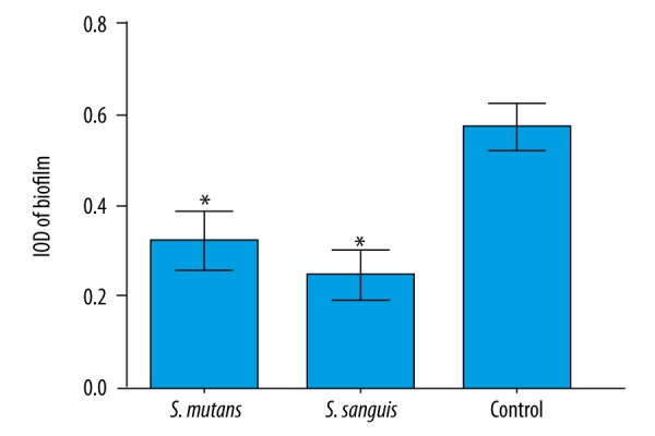
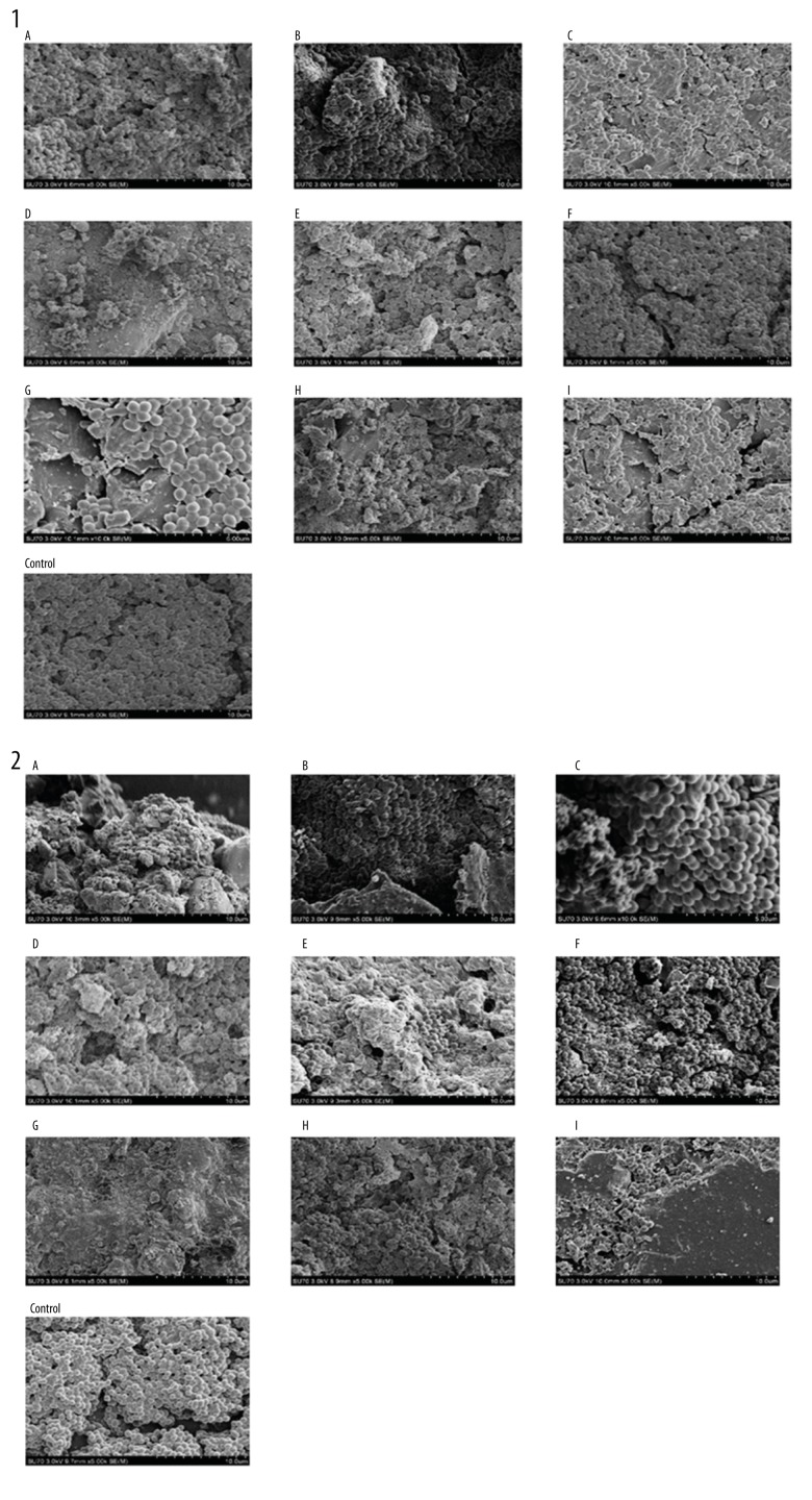
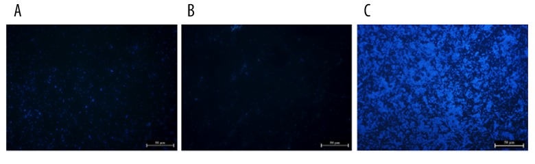
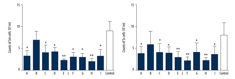
BACKGROUND Caries and periodontal diseases are caused by the biofilm formed by caries- and periodontal disease-related bacteria. Specific biofilms could be formed on different filling materials in oral cavity. Thus, to explore the inhibition effect of restorative filling materials on biofilm formation is of great significance in the treatment of caries and periodontal disease. MATERIAL AND METHODS The supernatants of S. mutans, S. sanguinis, and P. gingivalis suspension were combined with BHI broth. After 24 h, the live P. gingivalis number was calculated by colony counting and the biofilm was monitored by fluorescence microscopy. To test the adhesive ability of S. mutans and S. sanguinis on different dental materials, the biofilm was formed on different dental materials and then the bacterial number was calculated by using a Spectramax 250 microplate reader at OD 550, and the adhesive ability of S. mutans and S. sanguinis on different dental materials was analyzed by scanning electron microscopy. RESULTS The growth and biofilm formation of P. gingivalis was significantly inhibited by S. mutans and S. sanguinis supernatants (P<0.05). All groups except the zinc phosphate cement group (B) exerted a strong inhibitory effect on the biofilm formation of S. mutans and S. sanguinis (P<0.05). CONCLUSIONS The supernatants of S. mutans and S. sanguinis significantly inhibited the growth and biofilm formation of P. gingivalis, and the adhesive ability of S. mutans and S. sanguinis are different on different dental materials. These results provide useful information on dental caries, periodontal disease, and dental materials.
龋病和牙周病是由致龋菌和牙周病相关细菌形成的生物膜引起的。特定的生物膜可以在口腔中的不同填充材料上形成。因此,探索修复填充材料对生物膜形成的抑制作用对于龋病和牙周病的治疗具有重要意义。
将变形链球菌、血链球菌和牙龈卟啉单胞菌悬浮液的上清液与 BHI 肉汤混合。24 h 后,通过平板计数法计算活牙龈卟啉单胞菌数量,并通过荧光显微镜监测生物膜。为了测试变形链球菌和血链球菌在不同牙科材料上的粘附能力,在不同牙科材料上形成生物膜,然后使用 Spectramax 250 微孔板读数器在 OD550 处计算细菌数量,并通过扫描电子显微镜分析变形链球菌和血链球菌在不同牙科材料上的粘附能力。
变形链球菌和血链球菌上清液显著抑制牙龈卟啉单胞菌的生长和生物膜形成(P<0.05)。除磷酸锌水门汀组(B)外,所有组均对变形链球菌和血链球菌生物膜形成有很强的抑制作用(P<0.05)。
变形链球菌和血链球菌的上清液显著抑制牙龈卟啉单胞菌的生长和生物膜形成,变形链球菌和血链球菌在不同牙科材料上的粘附能力不同。这些结果为龋齿、牙周病和牙科材料提供了有用的信息。