Shafi Saba, Kellough David A, Lujan Giovanni, Satturwar Swati, Parwani Anil V, Li Zaibo
Department of Pathology, Wexner Medical Center at The Ohio State University, 410 W. 10th Ave, Columbus, OH 43210, USA.
J Pathol Inform. 2022 Jun 30;13:100122. doi: 10.1016/j.jpi.2022.100122. eCollection 2022.
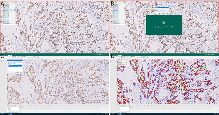
The Visiopharm automated estrogen receptor (ER) digital imaging analysis (DIA) algorithm assesses digitized ER immunohistochemistry (IHC) by segmenting tumor nuclei and detecting stained nuclei automatically. We aimed to integrate and validate this algorithm in a digital pathology workflow for clinical use.
The study cohort consisted of a serial collection of 97 invasive breast carcinoma specimens including 73 biopsies and 24 resections. ER IHC slides were scanned into Philips Image Management System (IMS) during our routine digital workflow and digital images were directly streamed into Visiopharm platform and analyzed using automated ER algorithm to obtain the positively stained tumor nuclei and staining intensity. ER DIA scores were compared with pathologists' manual scores.
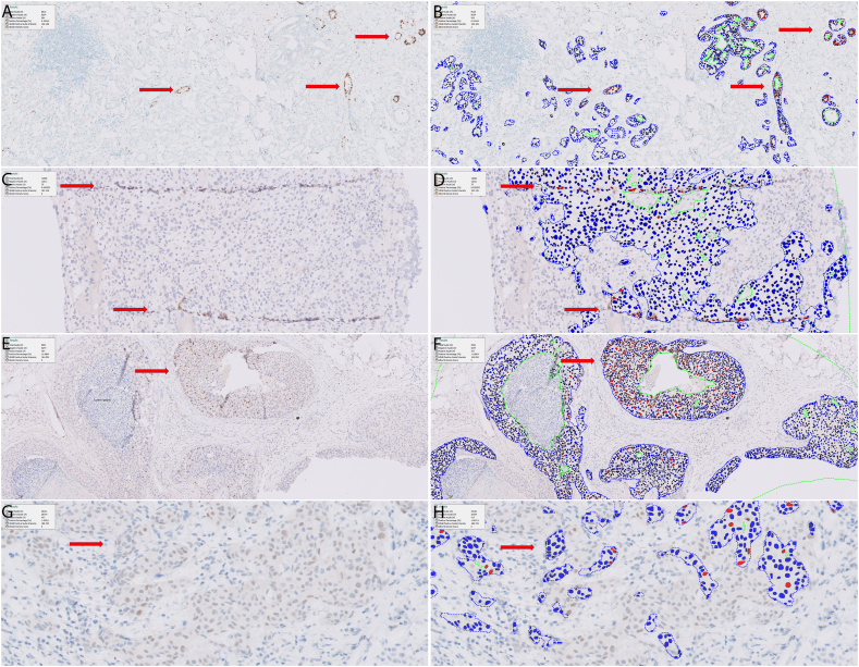
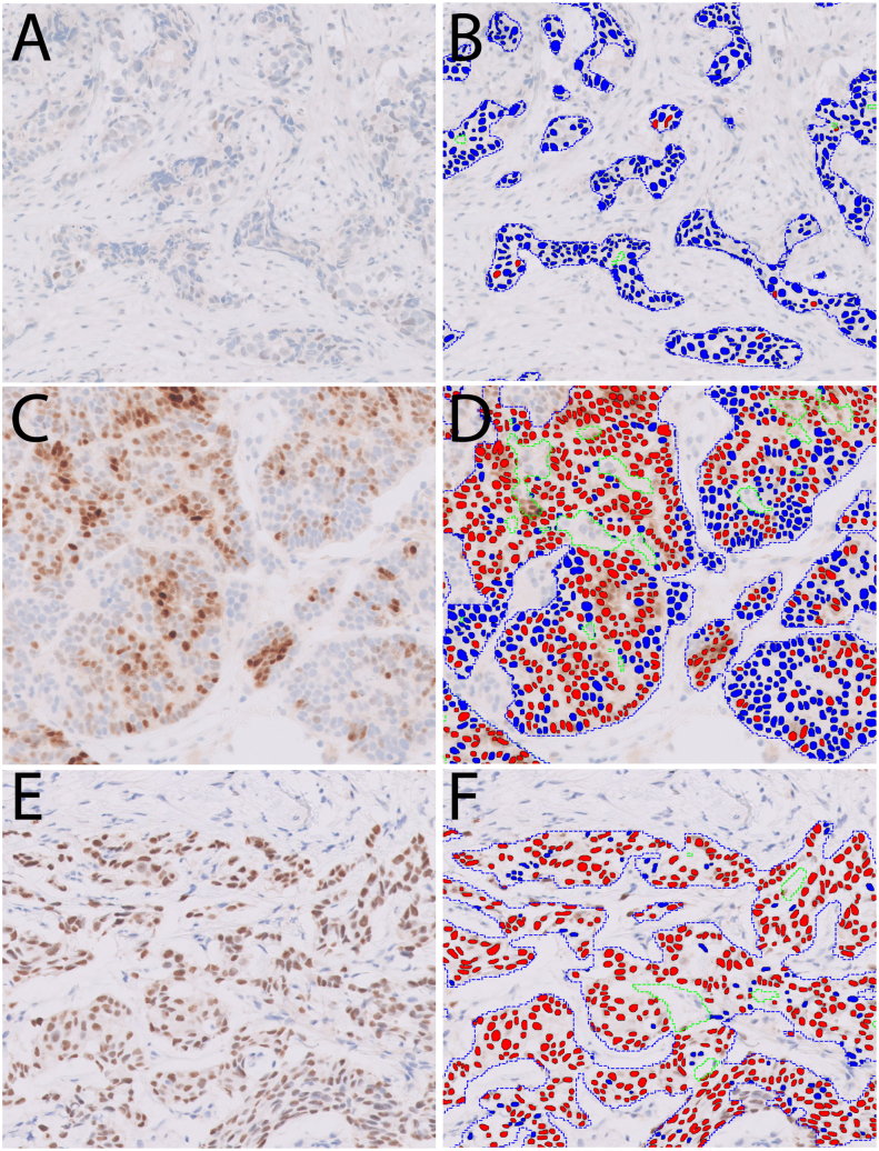
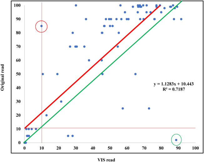
The overall concordance between pathologists' reads and DIA reads was excellent (91/97, 93.8%). Pearson Correlation Coefficient of the percentage of ER positive nuclei between the original reads and VIS reads was 0.72. Six cases (3 ER-negative and 3 ER-positive) had discordant results. All 3 false negative cases had very weak ER staining and no more than 10% positivity. The causes for false positive DIA were mainly pre-analytic/pre-imaging and included intermixed benign glands in tumor area, ductal carcinoma in-situ (DCIS) components, and tissue folding.
Automated ER DIA demonstrates excellent concordance with pathologists' scores and accurately discriminates ER positive from negative cases. Furthermore, integrating automated biomarker DIA into a busy clinical digital workflow is feasible and may save time and labor for pathologists.
Visiopharm 自动雌激素受体(ER)数字成像分析(DIA)算法通过自动分割肿瘤细胞核并检测染色细胞核来评估数字化的 ER 免疫组织化学(IHC)。我们旨在将该算法整合并验证到用于临床的数字病理工作流程中。
研究队列包括连续收集的 97 例浸润性乳腺癌标本,其中包括 73 例活检标本和 24 例切除标本。在我们的常规数字工作流程中,将 ER IHC 玻片扫描到飞利浦图像管理系统(IMS)中,数字图像直接传输到 Visiopharm 平台,并使用自动 ER 算法进行分析,以获得阳性染色的肿瘤细胞核和染色强度。将 ER DIA 评分与病理学家的手动评分进行比较。
病理学家的读数与 DIA 读数之间的总体一致性非常好(91/97,93.8%)。原始读数与 VIS 读数之间 ER 阳性细胞核百分比的 Pearson 相关系数为 0.72。有 6 例(3 例 ER 阴性和 3 例 ER 阳性)结果不一致。所有 3 例假阴性病例的 ER 染色都非常弱,阳性率不超过 10%。DIA 假阳性的原因主要是分析前/成像前因素,包括肿瘤区域内混合的良性腺体、导管原位癌(DCIS)成分和组织折叠。
自动 ER DIA 与病理学家的评分具有很好的一致性,能够准确地区分 ER 阳性和阴性病例。此外,将自动生物标志物 DIA 整合到繁忙的临床数字工作流程中是可行的,并且可能为病理学家节省时间和劳动力。