• 文献检索
  • 文档翻译
  • 深度研究
  • 学术资讯
  • Suppr Zotero 插件Zotero 插件
  • 邀请有礼
  • 套餐&价格
  • 历史记录
应用&插件
Suppr Zotero 插件Zotero 插件浏览器插件Mac 客户端Windows 客户端微信小程序
定价
高级版会员购买积分包购买API积分包
服务
文献检索文档翻译深度研究API 文档MCP 服务
关于我们
关于 Suppr公司介绍联系我们用户协议隐私条款
关注我们

Suppr 超能文献

核心技术专利:CN118964589B侵权必究
粤ICP备2023148730 号-1Suppr @ 2026

文献检索

告别复杂PubMed语法,用中文像聊天一样搜索,搜遍4000万医学文献。AI智能推荐,让科研检索更轻松。

立即免费搜索

文件翻译

保留排版,准确专业,支持PDF/Word/PPT等文件格式,支持 12+语言互译。

免费翻译文档

深度研究

AI帮你快速写综述,25分钟生成高质量综述,智能提取关键信息,辅助科研写作。

立即免费体验

使用细胞的空间图对组织中的细胞间通讯进行建模。

Modeling intercellular communication in tissues using spatial graphs of cells.

机构信息

Institute of Computational Biology, Helmholtz Zentrum München, Neuherberg, Germany.

TUM School of Life Sciences Weihenstephan, Technical University of Munich, Freising, Germany.

出版信息

Nat Biotechnol. 2023 Mar;41(3):332-336. doi: 10.1038/s41587-022-01467-z. Epub 2022 Oct 27.

DOI:10.1038/s41587-022-01467-z
PMID:36302986
原文链接:https://pmc.ncbi.nlm.nih.gov/articles/PMC10017508/
Abstract

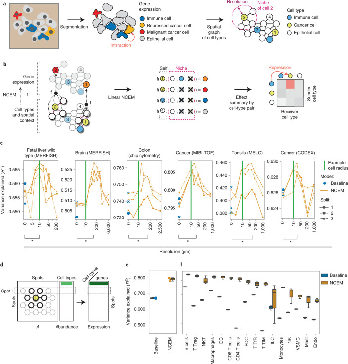
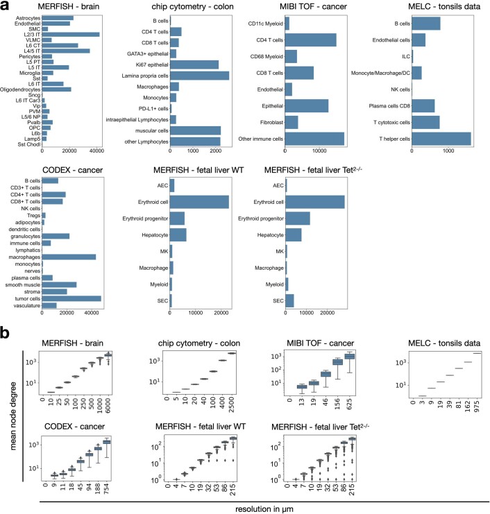
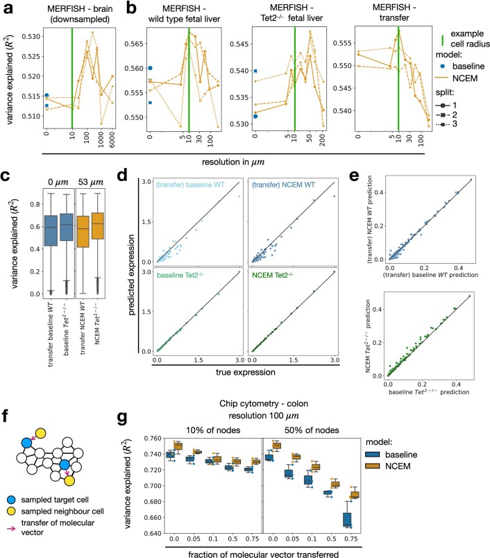
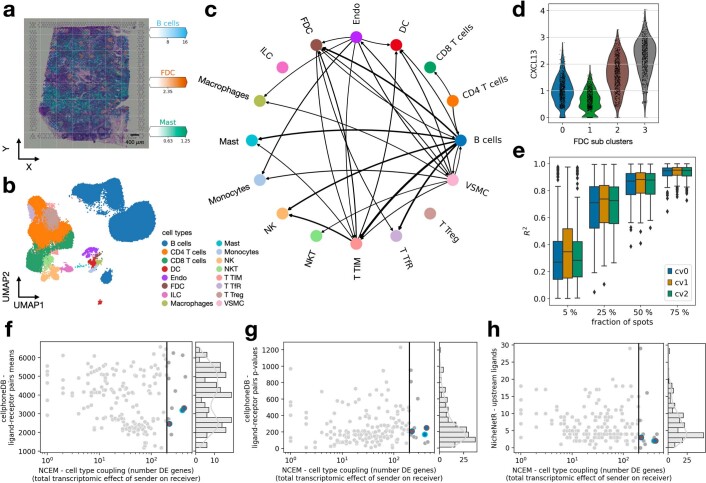
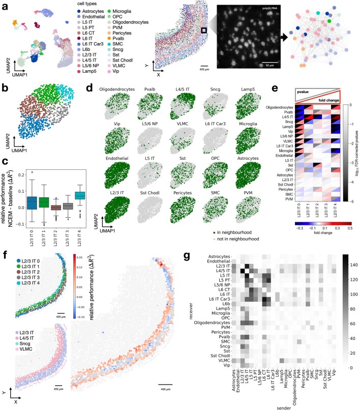
Models of intercellular communication in tissues are based on molecular profiles of dissociated cells, are limited to receptor-ligand signaling and ignore spatial proximity in situ. We present node-centric expression modeling, a method based on graph neural networks that estimates the effects of niche composition on gene expression in an unbiased manner from spatial molecular profiling data. We recover signatures of molecular processes known to underlie cell communication.

摘要

组织细胞间通讯模型基于分离细胞的分子谱,仅限于受体配体信号,并且忽略了原位的空间接近性。我们提出了以节点为中心的表达建模方法,该方法基于图神经网络,可以从空间分子分析数据中以无偏的方式估计生态位组成对基因表达的影响。我们恢复了已知是细胞通讯基础的分子过程的特征。

https://cdn.ncbi.nlm.nih.gov/pmc/blobs/6211/10017508/0ad7c6a83f14/41587_2022_1467_Fig12_ESM.jpg
https://cdn.ncbi.nlm.nih.gov/pmc/blobs/6211/10017508/6d6265140d62/41587_2022_1467_Fig1_HTML.jpg
https://cdn.ncbi.nlm.nih.gov/pmc/blobs/6211/10017508/2f9a094a80d4/41587_2022_1467_Fig2_HTML.jpg
https://cdn.ncbi.nlm.nih.gov/pmc/blobs/6211/10017508/3627f576f164/41587_2022_1467_Fig3_ESM.jpg
https://cdn.ncbi.nlm.nih.gov/pmc/blobs/6211/10017508/42f5e0a58246/41587_2022_1467_Fig4_ESM.jpg
https://cdn.ncbi.nlm.nih.gov/pmc/blobs/6211/10017508/e59314b1caa3/41587_2022_1467_Fig5_ESM.jpg
https://cdn.ncbi.nlm.nih.gov/pmc/blobs/6211/10017508/c683841349be/41587_2022_1467_Fig6_ESM.jpg
https://cdn.ncbi.nlm.nih.gov/pmc/blobs/6211/10017508/cfb40dce7e7e/41587_2022_1467_Fig7_ESM.jpg
https://cdn.ncbi.nlm.nih.gov/pmc/blobs/6211/10017508/e9a9242aa360/41587_2022_1467_Fig8_ESM.jpg
https://cdn.ncbi.nlm.nih.gov/pmc/blobs/6211/10017508/b4c01c2856a5/41587_2022_1467_Fig9_ESM.jpg
https://cdn.ncbi.nlm.nih.gov/pmc/blobs/6211/10017508/cd58f85bc1ca/41587_2022_1467_Fig10_ESM.jpg
https://cdn.ncbi.nlm.nih.gov/pmc/blobs/6211/10017508/a02c1addaa96/41587_2022_1467_Fig11_ESM.jpg
https://cdn.ncbi.nlm.nih.gov/pmc/blobs/6211/10017508/0ad7c6a83f14/41587_2022_1467_Fig12_ESM.jpg

相似文献

1
Modeling intercellular communication in tissues using spatial graphs of cells.使用细胞的空间图对组织中的细胞间通讯进行建模。
Nat Biotechnol. 2023 Mar;41(3):332-336. doi: 10.1038/s41587-022-01467-z. Epub 2022 Oct 27.
2
Specialized Intercellular Communications via Cytonemes and Nanotubes.细胞丝状伪足和纳米管的细胞间特异性通讯。
Annu Rev Cell Dev Biol. 2018 Oct 6;34:59-84. doi: 10.1146/annurev-cellbio-100617-062932. Epub 2018 Aug 3.
3
Exploring intercellular signaling by proteomic approaches.通过蛋白质组学方法探索细胞间信号传导。
Proteomics. 2014 Mar;14(4-5):498-512. doi: 10.1002/pmic.201300259. Epub 2013 Nov 11.
4
Explaining decisions of graph convolutional neural networks: patient-specific molecular subnetworks responsible for metastasis prediction in breast cancer.解释图卷积神经网络决策:乳腺癌转移预测中与患者特异性相关的分子子网络。
Genome Med. 2021 Mar 11;13(1):42. doi: 10.1186/s13073-021-00845-7.
5
Single-cell Transcriptome Profiling reveals Dermal and Epithelial cell fate decisions during Embryonic Hair Follicle Development.单细胞转录组谱分析揭示了胚胎毛囊发育过程中真皮和表皮细胞的命运决定。
Theranostics. 2020 Jun 12;10(17):7581-7598. doi: 10.7150/thno.44306. eCollection 2020.
6
Comparative analysis of cell-cell communication at single-cell resolution.单细胞分辨率下的细胞间通讯比较分析。
Nat Biotechnol. 2024 Mar;42(3):470-483. doi: 10.1038/s41587-023-01782-z. Epub 2023 May 11.
7
scTenifoldXct: A semi-supervised method for predicting cell-cell interactions and mapping cellular communication graphs.scTenifoldXct:一种用于预测细胞间相互作用和绘制细胞通信图谱的半监督方法。
Cell Syst. 2023 Apr 19;14(4):302-311.e4. doi: 10.1016/j.cels.2023.01.004. Epub 2023 Feb 13.
8
Dual graph convolutional neural network for predicting chemical networks.双图卷积神经网络用于预测化学网络。
BMC Bioinformatics. 2020 Apr 23;21(Suppl 3):94. doi: 10.1186/s12859-020-3378-0.
9
Graph Transformer Networks: Learning meta-path graphs to improve GNNs.图 Transformer 网络:学习元路径图以改进 GNNs。
Neural Netw. 2022 Sep;153:104-119. doi: 10.1016/j.neunet.2022.05.026. Epub 2022 Jun 4.
10
Kolmogorov complexity of epithelial pattern formation: the role of regulatory network configuration.上皮细胞模式形成的柯尔莫哥洛夫复杂性:调控网络配置的作用。
Biosystems. 2013 May;112(2):131-8. doi: 10.1016/j.biosystems.2013.03.005. Epub 2013 Mar 14.

引用本文的文献

1
Advancing biological understanding of cellular senescence with computational multiomics.利用计算多组学推进对细胞衰老的生物学理解。
Nat Genet. 2025 Sep 15. doi: 10.1038/s41588-025-02314-y.
2
Multimodal integration strategies for clinical application in oncology.肿瘤学临床应用中的多模态整合策略
Front Pharmacol. 2025 Aug 20;16:1609079. doi: 10.3389/fphar.2025.1609079. eCollection 2025.
3
Connectome-constrained ligand-receptor interaction analysis for understanding brain network communication.用于理解脑网络通信的连接体约束配体-受体相互作用分析

本文引用的文献

1
Multiplexed imaging and automated signal quantification in formalin-fixed paraffin-embedded tissues by ChipCytometry.通过 ChipCytometry 在福尔马林固定石蜡包埋组织中进行多重成像和自动化信号定量分析。
Cell Rep Methods. 2021 Oct 27;1(7):100104. doi: 10.1016/j.crmeth.2021.100104. eCollection 2021 Nov 22.
2
DestVI identifies continuums of cell types in spatial transcriptomics data.DestVI可识别空间转录组学数据中的细胞类型连续体。
Nat Biotechnol. 2022 Sep;40(9):1360-1369. doi: 10.1038/s41587-022-01272-8. Epub 2022 Apr 21.
3
Spatial components of molecular tissue biology.
Nat Commun. 2025 Sep 2;16(1):8179. doi: 10.1038/s41467-025-63204-9.
4
DECIPHER for learning disentangled cellular embeddings in large-scale heterogeneous spatial omics data.用于在大规模异质空间组学数据中学习解缠细胞嵌入的DECIPHER
Nat Commun. 2025 Aug 27;16(1):7991. doi: 10.1038/s41467-025-63140-8.
5
TrimNN: characterizing cellular community motifs for studying multicellular topological organization in complex tissues.TrimNN:表征细胞群落基序以研究复杂组织中的多细胞拓扑组织
Nat Commun. 2025 Aug 19;16(1):7737. doi: 10.1038/s41467-025-63141-7.
6
Systematic assessment of microenvironment-dependent transcriptional patterns and intercellular communication.对微环境依赖性转录模式和细胞间通讯的系统评估。
Genome Biol. 2025 Jul 6;26(1):193. doi: 10.1186/s13059-025-03677-5.
7
MEBOCOST maps metabolite-mediated intercellular communications using single-cell RNA-seq.MEBOCOST利用单细胞RNA测序绘制代谢物介导的细胞间通讯图谱。
Nucleic Acids Res. 2025 Jun 20;53(12). doi: 10.1093/nar/gkaf569.
8
QuadST identifies cell-cell interaction-changed genes in spatially resolved transcriptomics data.QuadST可在空间分辨转录组学数据中识别细胞间相互作用改变的基因。
Genome Res. 2025 Aug 1;35(8):1821-1831. doi: 10.1101/gr.279859.124.
9
Single-Cell RNA Sequencing Delineates Renal Anti-Fibrotic Mechanisms Mediated by TRPC6 Inhibition.单细胞RNA测序揭示TRPC6抑制介导的肾脏抗纤维化机制
Adv Sci (Weinh). 2025 Sep;12(33):e01175. doi: 10.1002/advs.202501175. Epub 2025 Jun 17.
10
Robust self-supervised machine learning for single cell embeddings and annotations.用于单细胞嵌入和注释的强大自监督机器学习
bioRxiv. 2025 Jun 8:2025.06.05.658097. doi: 10.1101/2025.06.05.658097.
分子组织生物学的空间组成部分。
Nat Biotechnol. 2022 Mar;40(3):308-318. doi: 10.1038/s41587-021-01182-1. Epub 2022 Feb 7.
4
Squidpy: a scalable framework for spatial omics analysis.鱿鱼皮:一种用于空间组学分析的可扩展框架。
Nat Methods. 2022 Feb;19(2):171-178. doi: 10.1038/s41592-021-01358-2. Epub 2022 Jan 31.
5
Cell2location maps fine-grained cell types in spatial transcriptomics.细胞定位图谱精细的细胞类型在空间转录组学。
Nat Biotechnol. 2022 May;40(5):661-671. doi: 10.1038/s41587-021-01139-4. Epub 2022 Jan 13.
6
Deep learning and alignment of spatially resolved single-cell transcriptomes with Tangram.基于 Tangram 的空间分辨单细胞转录组的深度学习和对齐。
Nat Methods. 2021 Nov;18(11):1352-1362. doi: 10.1038/s41592-021-01264-7. Epub 2021 Oct 28.
7
Integration of spatial and single-cell transcriptomic data elucidates mouse organogenesis.空间转录组和单细胞转录组数据的整合揭示了小鼠器官发生。
Nat Biotechnol. 2022 Jan;40(1):74-85. doi: 10.1038/s41587-021-01006-2. Epub 2021 Sep 6.
8
Spatial transcriptome profiling by MERFISH reveals fetal liver hematopoietic stem cell niche architecture.通过MERFISH进行的空间转录组分析揭示了胎儿肝脏造血干细胞微环境结构。
Cell Discov. 2021 Jun 29;7(1):47. doi: 10.1038/s41421-021-00266-1.
9
Multiplexed histology analyses for the phenotypic and spatial characterization of human innate lymphoid cells.多指标组织学分析鉴定人类固有淋巴细胞的表型和空间特征。
Nat Commun. 2021 Mar 19;12(1):1737. doi: 10.1038/s41467-021-21994-8.
10
Giotto: a toolbox for integrative analysis and visualization of spatial expression data.Giotto:一个用于空间表达数据综合分析和可视化的工具包。
Genome Biol. 2021 Mar 8;22(1):78. doi: 10.1186/s13059-021-02286-2.