Data science of Bioimages Lab, Faculty of Medicine and University Hospital Cologne, Center for Molecular Medicine Cologne (CMMC), University of Cologne, 50931, Cologne, Germany.
Department of General, Visceral, Cancer and Transplantation Surgery, University of Cologne, 50937, Cologne, Germany.
Br J Cancer. 2023 Mar;128(7):1369-1376. doi: 10.1038/s41416-023-02143-y. Epub 2023 Jan 30.
Fast and accurate diagnostics are key for personalised medicine. Particularly in cancer, precise diagnosis is a prerequisite for targeted therapies, which can prolong lives. In this work, we focus on the automatic identification of gastroesophageal adenocarcinoma (GEA) patients that qualify for a personalised therapy targeting epidermal growth factor receptor 2 (HER2). We present a deep-learning method for scoring microscopy images of GEA for the presence of HER2 overexpression.
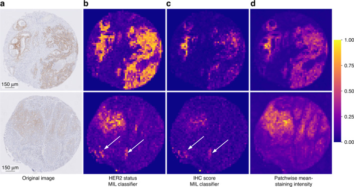
Our method is based on convolutional neural networks (CNNs) trained on a rich dataset of 1602 patient samples and tested on an independent set of 307 patient samples. We additionally verified the CNN's generalisation capabilities with an independent dataset with 653 samples from a separate clinical centre. We incorporated an attention mechanism in the network architecture to identify the tissue regions, which are important for the prediction outcome. Our solution allows for direct automated detection of HER2 in immunohistochemistry-stained tissue slides without the need for manual assessment and additional costly in situ hybridisation (ISH) tests.
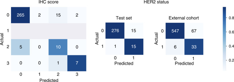
We show accuracy of 0.94, precision of 0.97, and recall of 0.95. Importantly, our approach offers accurate predictions in cases that pathologists cannot resolve and that require additional ISH testing. We confirmed our findings in an independent dataset collected in a different clinical centre. The attention-based CNN exploits morphological information in microscopy images and is superior to a predictive model based on the staining intensity only.
We demonstrate that our approach not only automates an important diagnostic process for GEA patients but also paves the way for the discovery of new morphological features that were previously unknown for GEA pathology.
快速准确的诊断是个性化医疗的关键。特别是在癌症中,精确诊断是靶向治疗的前提,靶向治疗可以延长生命。在这项工作中,我们专注于自动识别有资格接受针对表皮生长因子受体 2 (HER2) 的靶向治疗的胃食管腺癌 (GEA) 患者。我们提出了一种用于评分 GEA 显微镜图像中 HER2 过表达的深度学习方法。
我们的方法基于在包含 1602 个患者样本的丰富数据集上训练的卷积神经网络 (CNN),并在包含 307 个患者样本的独立数据集上进行测试。我们还使用来自另一个临床中心的包含 653 个样本的独立数据集验证了 CNN 的泛化能力。我们在网络架构中加入了注意力机制,以识别对预测结果重要的组织区域。我们的解决方案允许直接在免疫组织化学染色的组织切片中自动检测 HER2,而无需手动评估和额外的昂贵原位杂交 (ISH) 测试。
我们的准确率为 0.94,精确率为 0.97,召回率为 0.95。重要的是,我们的方法在病理学家无法解决且需要额外的 ISH 测试的情况下提供了准确的预测。我们在另一个临床中心收集的独立数据集中证实了我们的发现。基于注意力的 CNN 利用了显微镜图像中的形态学信息,优于仅基于染色强度的预测模型。
我们证明,我们的方法不仅自动化了 GEA 患者的重要诊断过程,而且为发现以前未知的 GEA 病理学新形态特征铺平了道路。