Gan Fan, Wu Fei-Peng, Zhong Yu-Lin
Medical College of Nanchang University, Nanchang, China.
Department of Ophthalmology, Jiangxi Provincial People's Hospital, The First Affiliated Hospital of Nanchang Medical College, Nanchang, China.
Front Neurosci. 2023 Jan 30;17:1097291. doi: 10.3389/fnins.2023.1097291. eCollection 2023.
A common ocular manifestation, macular edema (ME) is the primary cause of visual deterioration. In this study, an artificial intelligence method based on multi-feature fusion was introduced to enable automatic ME classification on spectral-domain optical coherence tomography (SD-OCT) images, to provide a convenient method of clinical diagnosis.
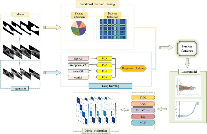
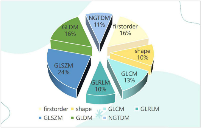
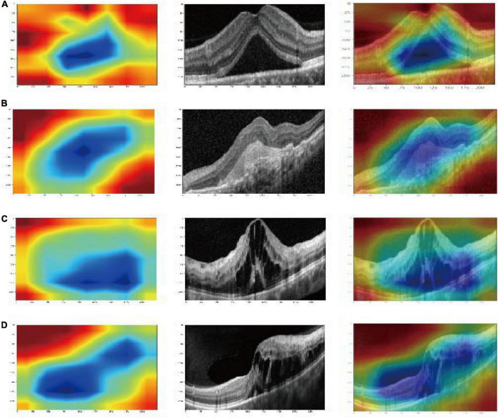
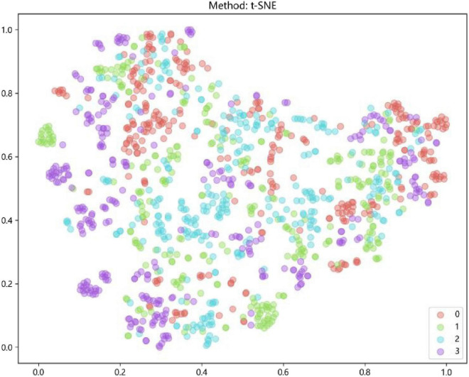
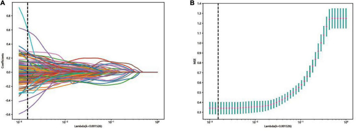
First, 1,213 two-dimensional (2D) cross-sectional OCT images of ME were collected from the Jiangxi Provincial People's Hospital between 2016 and 2021. According to OCT reports of senior ophthalmologists, there were 300 images with diabetic (DME), 303 images with age-related macular degeneration (AMD), 304 images with retinal-vein occlusion (RVO), and 306 images with central serous chorioretinopathy (CSC). Then, traditional omics features of the images were extracted based on the first-order statistics, shape, size, and texture. After extraction by the alexnet, inception_v3, resnet34, and vgg13 models and selected by dimensionality reduction using principal components analysis (PCA), the deep-learning features were fused. Next, the gradient-weighted class-activation map (Grad-CAM) was used to visualize the-deep-learning process. Finally, the fusion features set, which was fused from the traditional omics features and the deep-fusion features, was used to establish the final classification models. The performance of the final models was evaluated by accuracy, confusion matrix, and the receiver operating characteristic (ROC) curve.
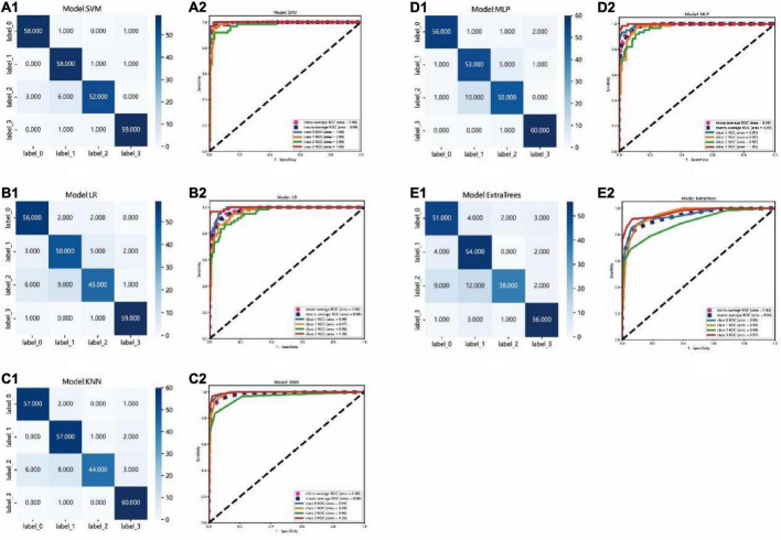
Compared with other classification models, the performance of the support vector machine (SVM) model was best, with an accuracy of 93.8%. The area under curves AUC of micro- and macro-averages were 99%, and the AUC of the AMD, DME, RVO, and CSC groups were 100, 99, 98, and 100%, respectively.
The artificial intelligence model in this study could be used to classify DME, AME, RVO, and CSC accurately from SD-OCT images.
黄斑水肿(ME)是一种常见的眼部表现,是视力下降的主要原因。本研究引入一种基于多特征融合的人工智能方法,以实现对光谱域光学相干断层扫描(SD-OCT)图像的ME自动分类,提供一种便捷的临床诊断方法。
首先,2016年至2021年期间从江西省人民医院收集了1213张ME的二维(2D)横断面OCT图像。根据高级眼科医生的OCT报告,其中有300张糖尿病性黄斑水肿(DME)图像、303张年龄相关性黄斑变性(AMD)图像、304张视网膜静脉阻塞(RVO)图像和306张中心性浆液性脉络膜视网膜病变(CSC)图像。然后,基于一阶统计、形状、大小和纹理提取图像的传统组学特征。经alexnet、inception_v3、resnet34和vgg13模型提取并通过主成分分析(PCA)进行降维选择后,融合深度学习特征。接下来,使用梯度加权类激活映射(Grad-CAM)对深度学习过程进行可视化。最后,将从传统组学特征和深度融合特征融合得到的融合特征集用于建立最终分类模型。通过准确率、混淆矩阵和受试者工作特征(ROC)曲线评估最终模型的性能。
与其他分类模型相比,支持向量机(SVM)模型性能最佳,准确率为93.8%。微观和宏观平均值的曲线下面积AUC为99%,AMD、DME、RVO和CSC组的AUC分别为100%、99%、98%和100%。
本研究中的人工智能模型可用于从SD-OCT图像中准确分类DME、AME、RVO和CSC。