School of Health & Life Sciences, Teesside University, TS1 3BX, Middlesbrough, UK.
National Horizons Centre, Teesside University, 38 John Dixon Ln, DL1 1HG, Darlington, UK.
BMC Cancer. 2023 Feb 21;23(1):174. doi: 10.1186/s12885-023-10588-w.
Gliomas are the most common brain tumours with the high-grade glioblastoma representing the most aggressive and lethal form. Currently, there is a lack of specific glioma biomarkers that would aid tumour subtyping and minimally invasive early diagnosis. Aberrant glycosylation is an important post-translational modification in cancer and is implicated in glioma progression. Raman spectroscopy (RS), a vibrational spectroscopic label-free technique, has already shown promise in cancer diagnostics.
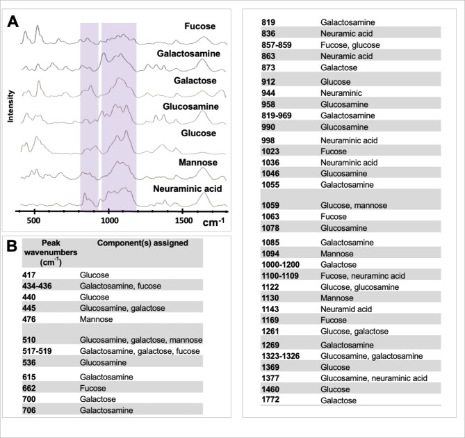
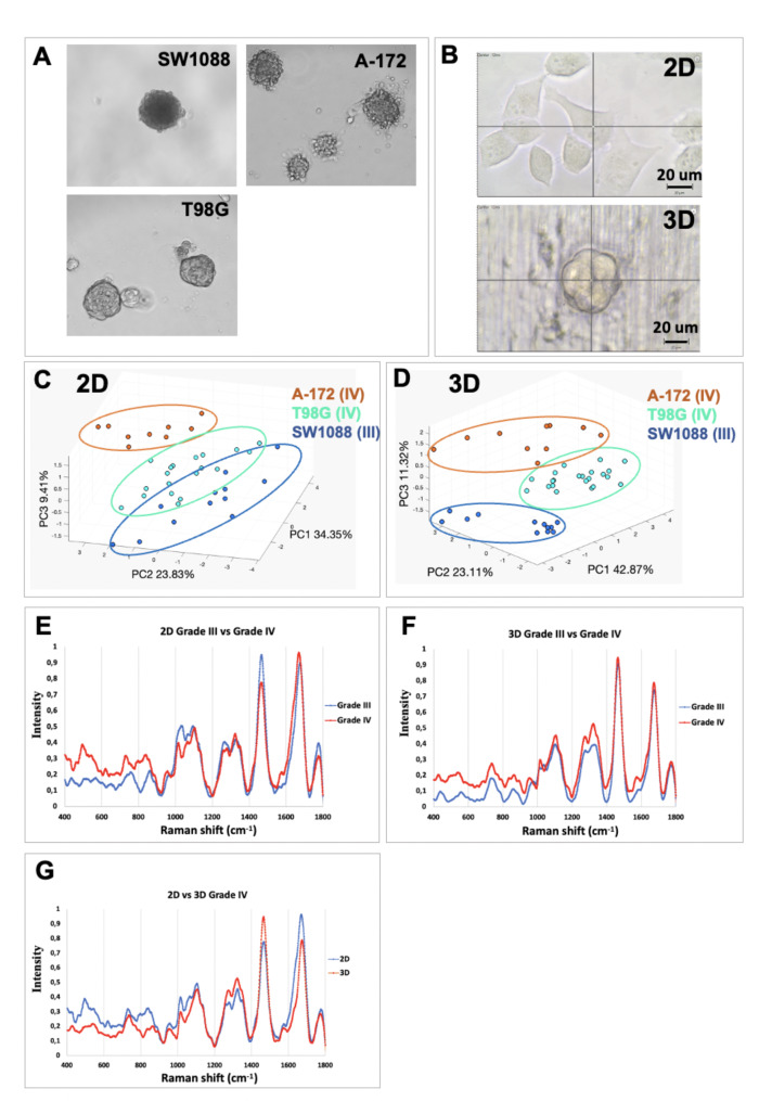
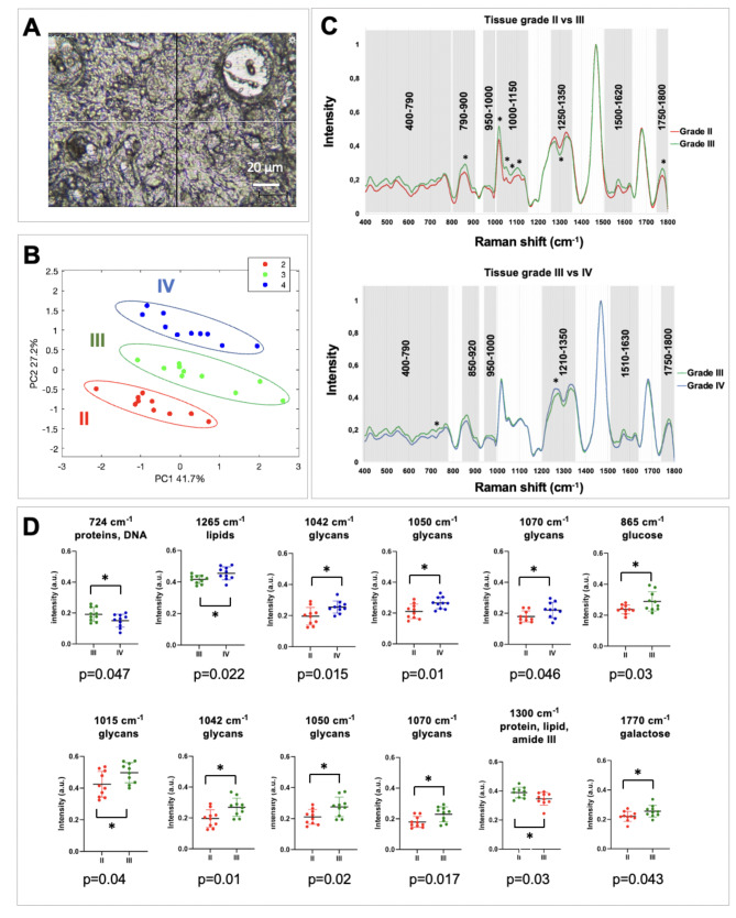
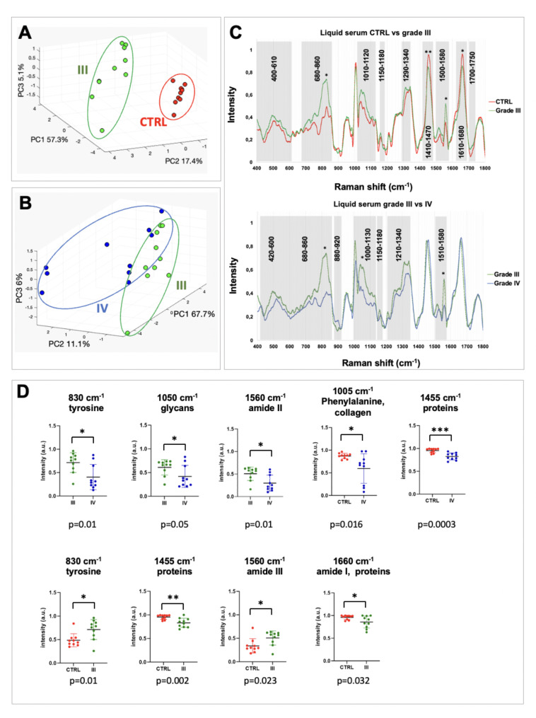
RS was combined with machine learning to discriminate glioma grades. Raman spectral signatures of glycosylation patterns were used in serum samples and fixed tissue biopsy samples, as well as in single cells and spheroids.
Glioma grades in fixed tissue patient samples and serum were discriminated with high accuracy. Discrimination between higher malignant glioma grades (III and IV) was achieved with high accuracy in tissue, serum, and cellular models using single cells and spheroids. Biomolecular changes were assigned to alterations in glycosylation corroborated by analysing glycan standards and other changes such as carotenoid antioxidant content.
RS combined with machine learning could pave the way for more objective and less invasive grading of glioma patients, serving as a useful tool to facilitate glioma diagnosis and delineate biomolecular glioma progression changes.
神经胶质瘤是最常见的脑肿瘤,高级别神经胶质瘤是最具侵袭性和致命性的形式。目前,缺乏特定的神经胶质瘤生物标志物来辅助肿瘤分型和微创早期诊断。糖基化异常是癌症中一种重要的翻译后修饰,与神经胶质瘤的进展有关。拉曼光谱(RS)是一种无标记的振动光谱技术,在癌症诊断中已经显示出了很好的效果。
RS 与机器学习相结合,以区分神经胶质瘤的等级。在血清样本和固定组织活检样本中,以及在单细胞和球体中,使用糖基化模式的拉曼光谱特征。
对固定组织患者样本和血清中的神经胶质瘤等级进行了高精度的区分。在组织、血清和细胞模型中,使用单细胞和球体,可以高精度地区分高级别神经胶质瘤(III 级和 IV 级)。通过分析糖胺聚糖标准品和其他变化,如类胡萝卜素抗氧化剂含量,证实了生物分子变化归因于糖基化的改变。
RS 与机器学习相结合,可以为神经胶质瘤患者的更客观和微创分级铺平道路,作为一种有用的工具,有助于神经胶质瘤的诊断和描绘生物分子神经胶质瘤的进展变化。