Lunenfeld-Tanenbaum Research Institute, Sinai Health System, 25 Orde St, Suite 6-1017, Toronto, ON, M5T 3H7, Canada.
Department of Physiology, University of Toronto, Toronto, Canada.
Sci Rep. 2023 Mar 23;13(1):4746. doi: 10.1038/s41598-023-31780-9.
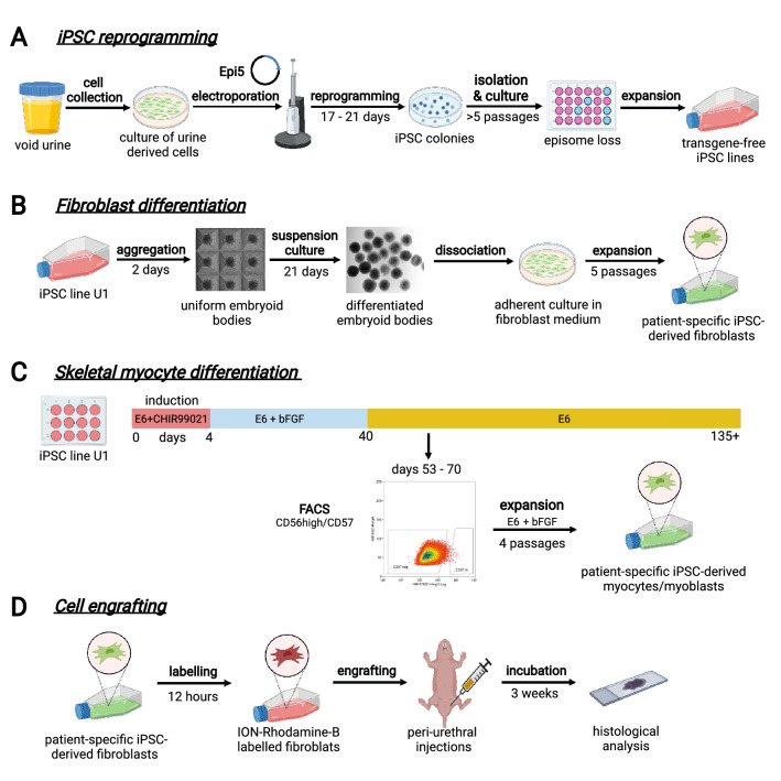
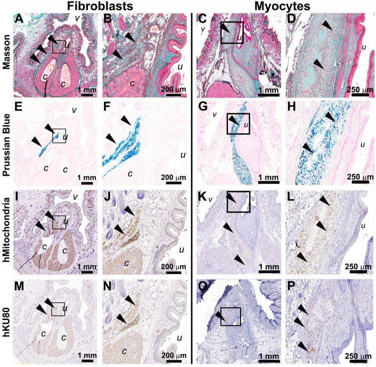
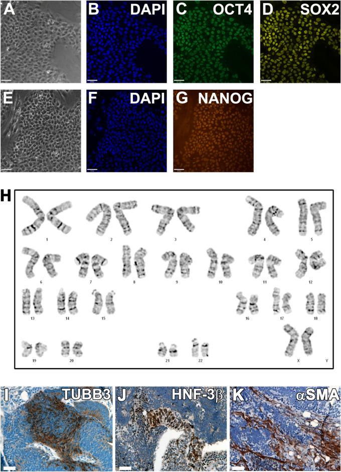
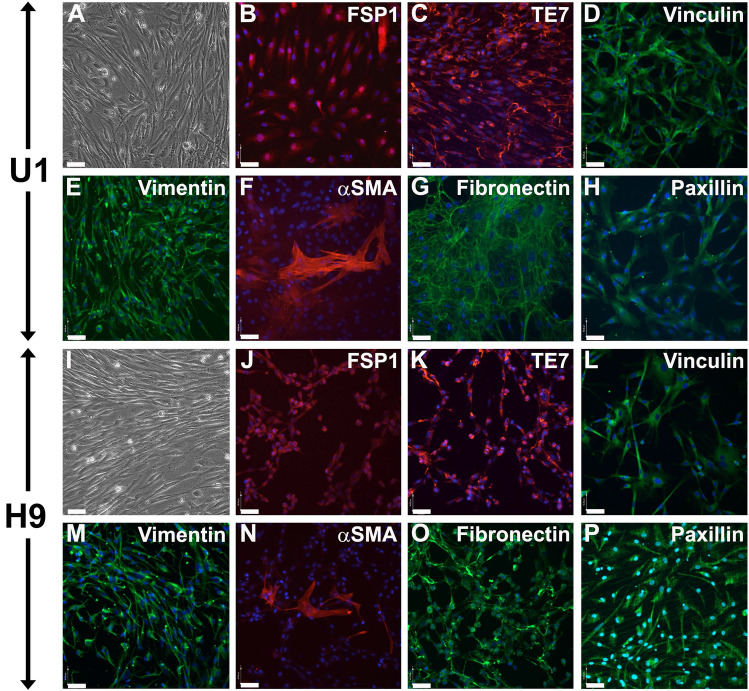
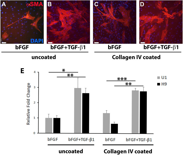
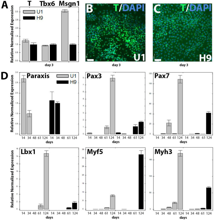
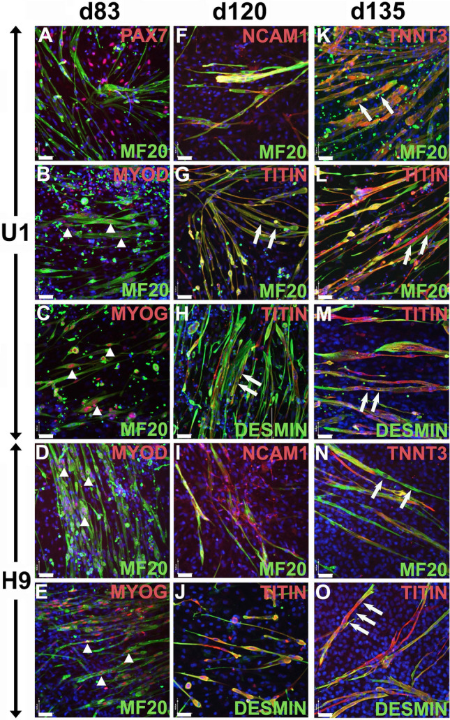
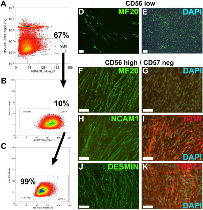
Cell-based therapy is a major focus for treatment of stress urinary incontinence (SUI). However, derivation of primary cells requires tissue biopsies, which often have adverse effects on patients. A recent study used human induced pluripotent stem cells (iPSC)-derived smooth muscle myocytes for urethral sphincter regeneration in rats. Here, we establish a workflow using iPSC-derived fibroblasts and skeletal myocytes for urethral tissue regeneration: (1) Cells from voided urine of women were reprogrammed into iPSC. (2) The iPSC line U1 and hESC line H9 (control) were differentiated into fibroblasts expressing FSP1, TE7, vinculin, vimentin, αSMA, fibronectin and paxillin. (3) Myogenic differentiation of U1 and H9 was induced by small molecule CHIR99021 and confirmed by protein expression of myogenic factors PAX7, MYOD, MYOG, and MF20. Striated muscle cells enriched by FACS expressed NCAM1, TITIN, DESMIN, TNNT3. (4) Human iPSC-derived fibroblasts and myocytes were engrafted into the periurethral region of RNU rats. Injected cells were labelled with ferric nanoparticles and traced by Prussian Blue stain, human-specific nuclear protein KU80, and human anti-mitochondria antibody. This workflow allows the scalable derivation, culture, and in vivo tracing of patient-specific fibroblasts and myocytes, which can be assessed in rat SUI models to regenerate urethral damages and restore continence.
基于细胞的疗法是治疗压力性尿失禁 (SUI) 的主要关注点。然而,原代细胞的衍生需要组织活检,这常常对患者产生不良影响。最近的一项研究使用人诱导多能干细胞 (iPSC) 衍生的平滑肌肌细胞在大鼠中进行尿道括约肌再生。在这里,我们建立了一个使用 iPSC 衍生的成纤维细胞和骨骼肌细胞进行尿道组织再生的工作流程:(1) 从女性的尿液中分离出细胞,将其重编程为 iPSC。(2) iPSC 系 U1 和 hESC 系 H9(对照)分化为表达 FSP1、TE7、波形蛋白、vimentin、αSMA、纤连蛋白和桩蛋白的成纤维细胞。(3) 通过小分子 CHIR99021 诱导 U1 和 H9 的肌生成分化,并通过肌生成因子 PAX7、MYOD、MYOG 和 MF20 的蛋白表达进行确认。通过 FACS 富集的横纹肌细胞表达 NCAM1、TITIN、DESMIN、TNNT3。(4) 将人 iPSC 衍生的成纤维细胞和肌细胞移植到 RNU 大鼠的尿道周围区域。注射的细胞用铁纳米颗粒标记,并通过普鲁士蓝染色、人特异性核蛋白 KU80 和人抗线粒体抗体进行追踪。该工作流程允许对患者特异性成纤维细胞和肌细胞进行可扩展的衍生、培养和体内追踪,可在大鼠 SUI 模型中评估这些细胞,以再生尿道损伤并恢复尿控。