Clinic of Radiology, University of Health Sciences Turkey, Prof. Dr. Cemil Taşcığlu City Hospital, İstanbul, Turkey.
Clinic of Radiation Oncology, University of Health Sciences Turkey, Prof. Dr. Cemil Taşcığlu City Hospital, İstanbul, Turkey.
Diagn Interv Radiol. 2023 May 31;29(3):460-468. doi: 10.4274/dir.2022.221335. Epub 2022 Dec 21.
This study aimed to evaluate the potential of machine learning-based models for predicting carcinogenic human papillomavirus (HPV) oncogene types using radiomics features from magnetic resonance imaging (MRI).
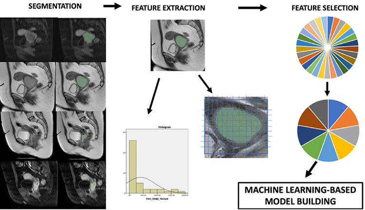
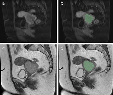
Pre-treatment MRI images of patients with cervical cancer were collected retrospectively. An HPV DNA oncogene analysis was performed based on cervical biopsy specimens. Radiomics features were extracted from contrast-enhanced T1-weighted images (CE-T1) and T2-weighted images (T2WI). A third feature subset was created as a combined group by concatenating the CE-T1 and T2WI subsets. Feature selection was performed using Pearson's correlation coefficient and wrapper- based sequential-feature selection. Two models were built with each feature subset, using support vector machine (SVM) and logistic regression (LR) classifiers. The models were validated using a five-fold cross-validation technique and compared using Wilcoxon's signed rank and Friedman's tests.
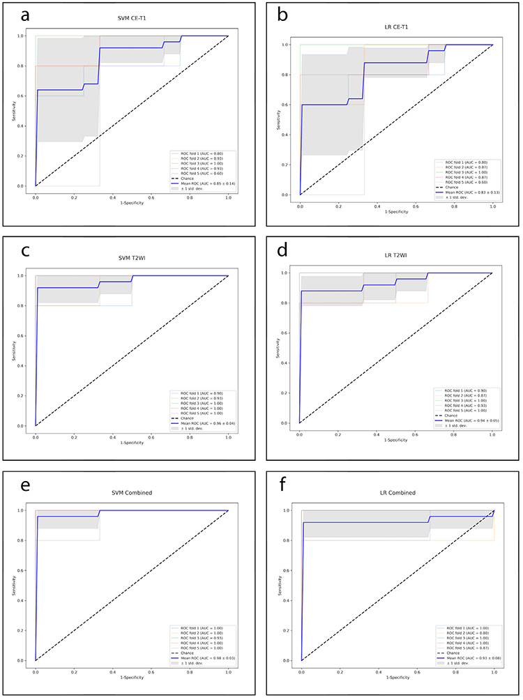
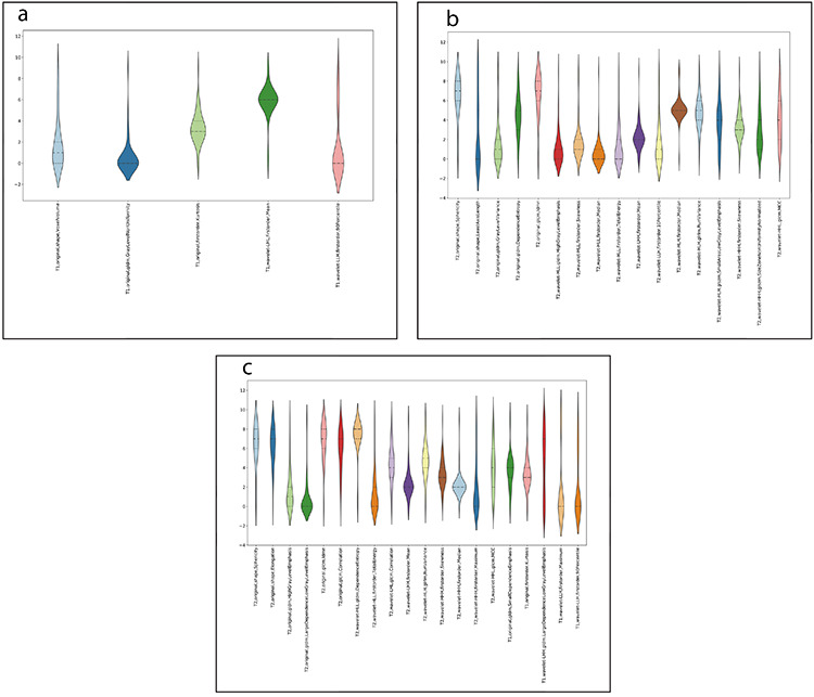
Forty-one patients were enrolled in the study (26 were positive for carcinogenic HPV oncogenes, and 15 were negative). A total of 851 features were extracted from each imaging sequence. After feature selection, 5, 17, and 20 features remained in the CE-T1, T2WI, and combined groups, respectively. The SVM models showed 83%, 95%, and 95% accuracy scores, and the LR models revealed 83%, 81%, and 92.5% accuracy scores in the CE-T1, T2WI, and combined groups, respectively. The SVM algorithm performed better than the LR algorithm in the T2WI feature subset ( = 0.005), and the feature sets in the T2WI and the combined group performed better than CE-T1 in the SVM model ( = 0.033 and 0.006, respectively). The combined group feature subset performed better than T2WI in the LR model ( = 0.023).
Machine learning-based radiomics models based on pre-treatment MRI can detect carcinogenic HPV status with discriminative accuracy.
本研究旨在评估基于机器学习的模型利用磁共振成像(MRI)的放射组学特征预测致癌性人乳头瘤病毒(HPV)致癌基因类型的潜力。
回顾性收集宫颈癌患者的预处理 MRI 图像。根据宫颈活检标本进行 HPV DNA 致癌基因分析。从对比增强 T1 加权图像(CE-T1)和 T2 加权图像(T2WI)中提取放射组学特征。第三组特征子集是通过串联 CE-T1 和 T2WI 子集创建的。使用 Pearson 相关系数和基于包装的顺序特征选择进行特征选择。使用支持向量机(SVM)和逻辑回归(LR)分类器为每个特征子集构建两个模型。使用五折交叉验证技术验证模型,并使用 Wilcoxon 符号秩和 Friedman 检验进行比较。
本研究纳入 41 例患者(26 例致癌 HPV 致癌基因阳性,15 例阴性)。每个成像序列共提取 851 个特征。经过特征选择,CE-T1、T2WI 和联合组中分别保留了 5、17 和 20 个特征。SVM 模型在 CE-T1、T2WI 和联合组中的准确率分别为 83%、95%和 95%,LR 模型的准确率分别为 83%、81%和 92.5%。SVM 算法在 T2WI 特征子集中的性能优于 LR 算法(=0.005),T2WI 和联合组中的特征集在 SVM 模型中的性能优于 CE-T1(分别为=0.033 和 0.006)。LR 模型中联合组特征子集的性能优于 T2WI(=0.023)。
基于预处理 MRI 的基于机器学习的放射组学模型可以以有区别的准确性检测致癌性 HPV 状态。