Institute of Biomedical Engineering, University of Toronto, Toronto, ON M5S 3G9, Canada.
Toronto General Research Institute, University Health Network, Toronto, ON M5G 2C4, Canada.
Biofabrication. 2023 Jun 22;15(3):035023. doi: 10.1088/1758-5090/acd8f4.
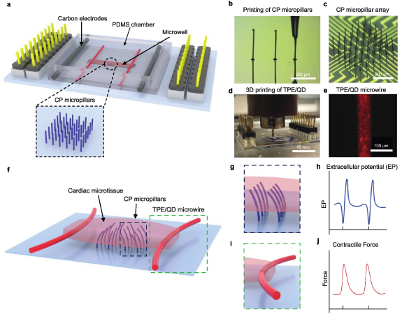
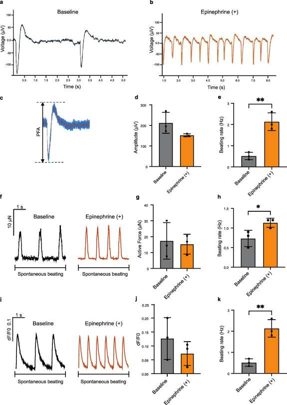
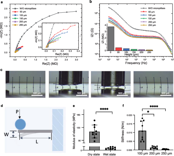
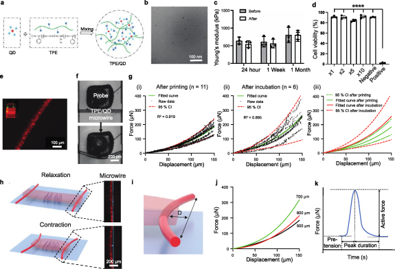
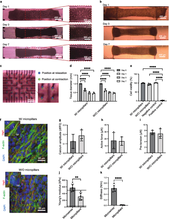
We developed a heart-on-a-chip platform that integrates highly flexible, vertical, 3D micropillar electrodes for electrophysiological recording and elastic microwires for the tissue's contractile force assessment. The high aspect ratio microelectrodes were 3D-printed into the device using a conductive polymer, poly(3,4-ethylenedioxythiophene):poly(styrene sulfonate) (PEDOT:PSS). A pair of flexible, quantum dots/thermoplastic elastomer nanocomposite microwires were 3D printed to anchor the tissue and enable continuous contractile force assessment. The 3D microelectrodes and flexible microwires enabled unobstructed human iPSC-based cardiac tissue formation and contraction, suspended above the device surface, under both spontaneous beating and upon pacing with a separate set of integrated carbon electrodes. Recording of extracellular field potentials using the PEDOT:PSS micropillars was demonstrated with and without epinephrine as a model drug, non-invasively, along withmonitoring of tissue contractile properties and calcium transients. Uniquely, the platform provides integrated profiling of electrical and contractile tissue properties, which is critical for proper evaluation of complex, mechanically and electrically active tissues, such as the heart muscle under both physiological and pathological conditions.
我们开发了一种芯片上的心脏模型平台,该平台集成了高度灵活的垂直 3D 微柱电极,用于电生理记录,以及弹性微丝,用于评估组织的收缩力。高纵横比微电极使用导电聚合物聚(3,4-亚乙基二氧噻吩):聚(苯乙烯磺酸盐)(PEDOT:PSS)3D 打印到器件中。一对灵活的量子点/热塑性弹性体纳米复合材料微丝被 3D 打印出来,以固定组织并实现连续的收缩力评估。3D 微电极和灵活的微丝使得在独立的集成碳电极起搏和自发跳动下,未被阻挡的基于人诱导多能干细胞的心脏组织能够在器件表面上方形成和收缩。使用 PEDOT:PSS 微柱记录细胞外场电势,同时使用和不使用肾上腺素作为模型药物进行记录,非侵入性监测组织收缩特性和钙瞬变。该平台的独特之处在于提供了电和收缩组织特性的综合分析,这对于正确评估复杂的、机械和电活性组织(如在生理和病理条件下的心肌)至关重要。