Nishimoto Soh, Saito Takuya, Ishise Hisako, Fujiwara Toshihiro, Kawai Kenichiro, Kakibuchi Masao
Department of Plastic Surgery, Hyogo Medical University, Nishinomiya 663-8501, Japan.
Diagnostics (Basel). 2023 Jun 1;13(11):1930. doi: 10.3390/diagnostics13111930.
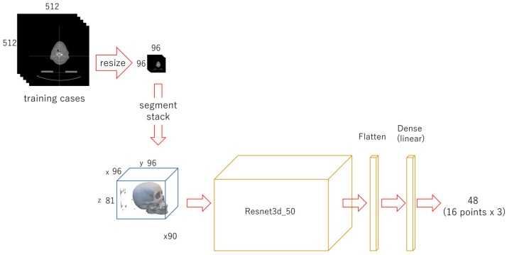
Geometrical assessments of human skulls have been conducted based on anatomical landmarks. If developed, the automatic detection of these landmarks will yield both medical and anthropological benefits. In this study, an automated system with multi-phased deep learning networks was developed to predict the three-dimensional coordinate values of craniofacial landmarks. Computed tomography images of the craniofacial area were obtained from a publicly available database. They were digitally reconstructed into three-dimensional objects. Sixteen anatomical landmarks were plotted on each of the objects, and their coordinate values were recorded. Three-phased regression deep learning networks were trained using ninety training datasets. For the evaluation, 30 testing datasets were employed. The 3D error for the first phase, which tested 30 data, was 11.60 px on average (1 px = 500/512 mm). For the second phase, it was significantly improved to 4.66 px. For the third phase, it was further significantly reduced to 2.88. This was comparable to the gaps between the landmarks, as plotted by two experienced practitioners. Our proposed method of multi-phased prediction, which conducts coarse detection first and narrows down the detection area, may be a possible solution to prediction problems, taking into account the physical limitations of memory and computation.
基于解剖学标志点对人类头骨进行了几何评估。如果能够实现这些标志点的自动检测,将在医学和人类学方面带来益处。在本研究中,开发了一个具有多阶段深度学习网络的自动化系统,用于预测颅面标志点的三维坐标值。从一个公开可用的数据库中获取颅面部区域的计算机断层扫描图像。将它们数字重建为三维物体。在每个物体上绘制了16个解剖学标志点,并记录了它们的坐标值。使用90个训练数据集对三阶段回归深度学习网络进行训练。为了进行评估,采用了30个测试数据集。测试30个数据的第一阶段的三维误差平均为11.60像素(1像素 = 500/512毫米)。第二阶段显著提高到4.66像素。第三阶段进一步显著降低到2.88像素。这与两位经验丰富的从业者绘制的标志点之间的间距相当。我们提出的多阶段预测方法,即先进行粗略检测并缩小检测区域,考虑到内存和计算的物理限制,可能是解决预测问题的一种可行方案。