To Tyrell, Lu Tongtong, Jorns Julie M, Patton Mollie, Schmidt Taly Gilat, Yen Tina, Yu Bing, Ye Dong Hye
Department of Electrical and Computer Engineering, Marquette University, Opus College of Engineering, Milwaukee, WI, United States.
Joint Department of Biomedical Engineering, Marquette University and Medical College of Wisconsin, Milwaukee, WI, United States.
Front Oncol. 2023 Jun 16;13:1179025. doi: 10.3389/fonc.2023.1179025. eCollection 2023.
Breast-conserving surgery is aimed at removing all cancerous cells while minimizing the loss of healthy tissue. To ensure a balance between complete resection of cancer and preservation of healthy tissue, it is necessary to assess themargins of the removed specimen during the operation. Deep ultraviolet (DUV) fluorescence scanning microscopy provides rapid whole-surface imaging (WSI) of resected tissues with significant contrast between malignant and normal/benign tissue. Intra-operative margin assessment with DUV images would benefit from an automated breast cancer classification method.
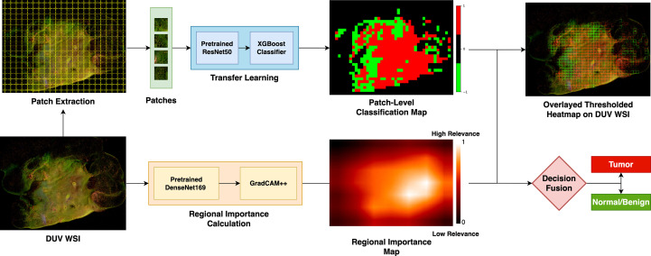
Deep learning has shown promising results in breast cancer classification, but the limited DUV image dataset presents the challenge of overfitting to train a robust network. To overcome this challenge, the DUV-WSI images are split into small patches, and features are extracted using a pre-trained convolutional neural network-afterward, a gradient-boosting tree trains on these features for patch-level classification. An ensemble learning approach merges patch-level classification results and regional importance to determine the margin status. An explainable artificial intelligence method calculates the regional importance values.
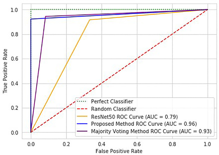
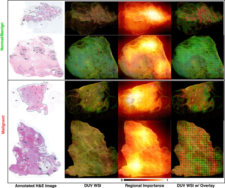
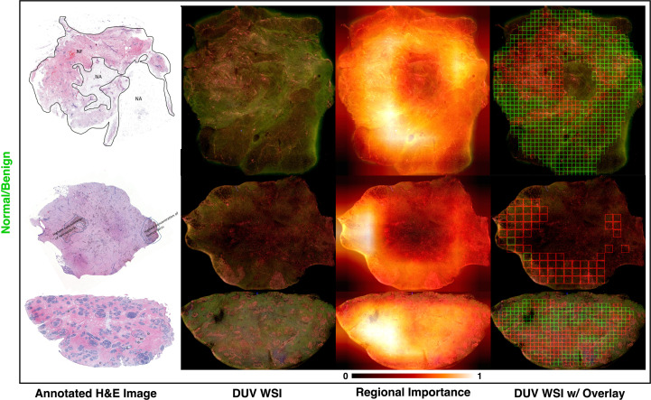
The proposed method's ability to determine the DUV WSI was high with 95% accuracy. The 100% sensitivity shows that the method can detect malignant cases efficiently. The method could also accurately localize areas that contain malignant or normal/benign tissue.
The proposed method outperforms the standard deep learning classification methods on the DUV breast surgical samples. The results suggest that it can be used to improve classification performance and identify cancerous regions more effectively.
保乳手术旨在切除所有癌细胞,同时尽量减少健康组织的损失。为确保在癌症的完整切除与健康组织的保留之间取得平衡,在手术过程中评估切除标本的切缘是必要的。深紫外(DUV)荧光扫描显微镜可对切除组织进行快速全表面成像(WSI),恶性组织与正常/良性组织之间具有显著对比度。使用DUV图像进行术中切缘评估将受益于一种自动乳腺癌分类方法。
深度学习在乳腺癌分类方面已显示出有前景的结果,但有限的DUV图像数据集给训练一个强大的网络带来了过拟合的挑战。为克服这一挑战,将DUV-WSI图像分割成小补丁,并使用预训练的卷积神经网络提取特征,之后,梯度提升树基于这些特征进行补丁级分类训练。一种集成学习方法合并补丁级分类结果和区域重要性以确定切缘状态。一种可解释人工智能方法计算区域重要性值。
所提出的方法确定DUV WSI的能力很高,准确率达95%。100%的灵敏度表明该方法能够有效地检测出恶性病例。该方法还能够准确地定位包含恶性或正常/良性组织的区域。
所提出的方法在DUV乳腺手术样本上优于标准的深度学习分类方法。结果表明它可用于提高分类性能并更有效地识别癌性区域。