• 文献检索
  • 文档翻译
  • 深度研究
  • 学术资讯
  • Suppr Zotero 插件Zotero 插件
  • 邀请有礼
  • 套餐&价格
  • 历史记录
应用&插件
Suppr Zotero 插件Zotero 插件浏览器插件Mac 客户端Windows 客户端微信小程序
定价
高级版会员购买积分包购买API积分包
服务
文献检索文档翻译深度研究API 文档MCP 服务
关于我们
关于 Suppr公司介绍联系我们用户协议隐私条款
关注我们

Suppr 超能文献

核心技术专利:CN118964589B侵权必究
粤ICP备2023148730 号-1Suppr @ 2026

文献检索

告别复杂PubMed语法,用中文像聊天一样搜索,搜遍4000万医学文献。AI智能推荐,让科研检索更轻松。

立即免费搜索

文件翻译

保留排版,准确专业,支持PDF/Word/PPT等文件格式,支持 12+语言互译。

免费翻译文档

深度研究

AI帮你快速写综述,25分钟生成高质量综述,智能提取关键信息,辅助科研写作。

立即免费体验

使用U-Net和深度卷积神经网络对CT扫描的卵巢肿瘤进行分割和分类的性能分析

Performance Analysis of Segmentation and Classification of CT-Scanned Ovarian Tumours Using U-Net and Deep Convolutional Neural Networks.

作者信息

Kodipalli Ashwini, Fernandes Steven L, Gururaj Vaishnavi, Varada Rameshbabu Shriya, Dasar Santosh

机构信息

Department of Artificial Intelligence & Data Science, Global Academy of Technology, Bangalore 560098, India.

Department of Computer Science, Design, Journalism, Creighton University, Omaha, NE 68178, USA.

出版信息

Diagnostics (Basel). 2023 Jul 5;13(13):2282. doi: 10.3390/diagnostics13132282.

DOI:10.3390/diagnostics13132282
PMID:37443676
原文链接:https://pmc.ncbi.nlm.nih.gov/articles/PMC10341135/
Abstract

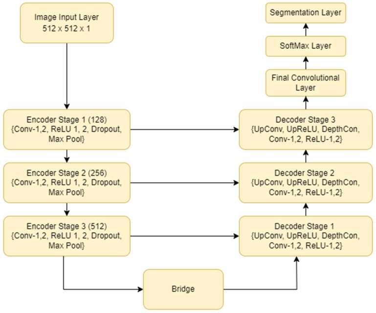
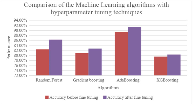
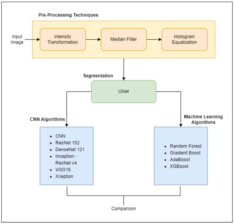
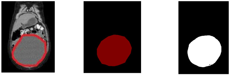
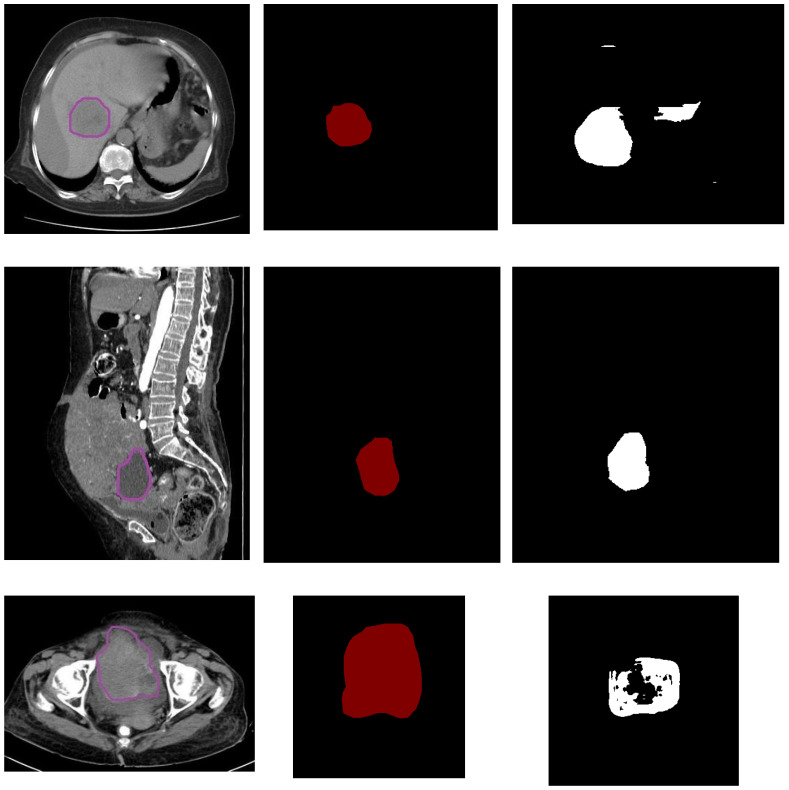
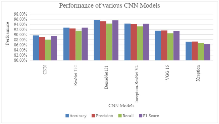
Difficulty in detecting tumours in early stages is the major cause of mortalities in patients, despite the advancements in treatment and research regarding ovarian cancer. Deep learning algorithms were applied to serve the purpose as a diagnostic tool and applied to CT scan images of the ovarian region. The images went through a series of pre-processing techniques and, further, the tumour was segmented using the UNet model. The instances were then classified into two categories-benign and malignant tumours. Classification was performed using deep learning models like CNN, ResNet, DenseNet, Inception-ResNet, VGG16 and Xception, along with machine learning models such as Random Forest, Gradient Boosting, AdaBoosting and XGBoosting. DenseNet 121 emerges as the best model on this dataset after applying optimization on the machine learning models by obtaining an accuracy of 95.7%. The current work demonstrates the comparison of multiple CNN architectures with common machine learning algorithms, with and without optimization techniques applied.

摘要

尽管在卵巢癌的治疗和研究方面取得了进展,但早期肿瘤难以检测仍是患者死亡的主要原因。深度学习算法被用作诊断工具,并应用于卵巢区域的CT扫描图像。这些图像经过了一系列预处理技术,然后使用UNet模型对肿瘤进行分割。接着将实例分为两类——良性肿瘤和恶性肿瘤。使用CNN、ResNet、DenseNet、Inception-ResNet、VGG16和Xception等深度学习模型以及随机森林、梯度提升、AdaBoosting和XGBoosting等机器学习模型进行分类。在对机器学习模型进行优化后,DenseNet 121在该数据集上成为最佳模型,准确率达到95.7%。当前的工作展示了在应用和未应用优化技术的情况下,多种CNN架构与常见机器学习算法的比较。

https://cdn.ncbi.nlm.nih.gov/pmc/blobs/63ff/10341135/d691a8ae62be/diagnostics-13-02282-g009.jpg
https://cdn.ncbi.nlm.nih.gov/pmc/blobs/63ff/10341135/48543e66a370/diagnostics-13-02282-g001.jpg
https://cdn.ncbi.nlm.nih.gov/pmc/blobs/63ff/10341135/ecdeda1267ef/diagnostics-13-02282-g002.jpg
https://cdn.ncbi.nlm.nih.gov/pmc/blobs/63ff/10341135/bc84693aa389/diagnostics-13-02282-g003a.jpg
https://cdn.ncbi.nlm.nih.gov/pmc/blobs/63ff/10341135/45c8affeb44d/diagnostics-13-02282-g004.jpg
https://cdn.ncbi.nlm.nih.gov/pmc/blobs/63ff/10341135/bd0ce40edf61/diagnostics-13-02282-g005.jpg
https://cdn.ncbi.nlm.nih.gov/pmc/blobs/63ff/10341135/86004b1d5a7b/diagnostics-13-02282-g006.jpg
https://cdn.ncbi.nlm.nih.gov/pmc/blobs/63ff/10341135/b64602e87993/diagnostics-13-02282-g007.jpg
https://cdn.ncbi.nlm.nih.gov/pmc/blobs/63ff/10341135/767fa3fefb25/diagnostics-13-02282-g008.jpg
https://cdn.ncbi.nlm.nih.gov/pmc/blobs/63ff/10341135/d691a8ae62be/diagnostics-13-02282-g009.jpg

相似文献

1
Performance Analysis of Segmentation and Classification of CT-Scanned Ovarian Tumours Using U-Net and Deep Convolutional Neural Networks.使用U-Net和深度卷积神经网络对CT扫描的卵巢肿瘤进行分割和分类的性能分析
Diagnostics (Basel). 2023 Jul 5;13(13):2282. doi: 10.3390/diagnostics13132282.
2
An Empirical Evaluation of a Novel Ensemble Deep Neural Network Model and Explainable AI for Accurate Segmentation and Classification of Ovarian Tumors Using CT Images.一种新型集成深度神经网络模型与可解释人工智能用于基于CT图像的卵巢肿瘤精确分割与分类的实证评估
Diagnostics (Basel). 2024 Mar 4;14(5):543. doi: 10.3390/diagnostics14050543.
3
Multiple skin lesions diagnostics via integrated deep convolutional networks for segmentation and classification.基于集成深度卷积网络的多皮肤损伤诊断,用于分割和分类。
Comput Methods Programs Biomed. 2020 Jul;190:105351. doi: 10.1016/j.cmpb.2020.105351. Epub 2020 Jan 23.
4
Automated detection of leukemia by pretrained deep neural networks and transfer learning: A comparison.基于预训练深度神经网络和迁移学习的白血病自动检测:比较研究。
Med Eng Phys. 2021 Dec;98:8-19. doi: 10.1016/j.medengphy.2021.10.006. Epub 2021 Oct 13.
5
Advancing maxillofacial prosthodontics by using pre-trained convolutional neural networks: Image-based classification of the maxilla.利用预训练卷积神经网络推进颌面赝复学:上颌骨的基于图像分类。
J Prosthodont. 2024 Aug;33(7):645-654. doi: 10.1111/jopr.13853. Epub 2024 Apr 3.
6
Deep learning for colon cancer histopathological images analysis.用于结肠癌组织病理学图像分析的深度学习
Comput Biol Med. 2021 Sep;136:104730. doi: 10.1016/j.compbiomed.2021.104730. Epub 2021 Aug 4.
7
Tongue image quality assessment based on a deep convolutional neural network.基于深度卷积神经网络的舌象质量评估。
BMC Med Inform Decis Mak. 2021 May 5;21(1):147. doi: 10.1186/s12911-021-01508-8.
8
Quantitative and Qualitative Analysis of 18 Deep Convolutional Neural Network (CNN) Models with Transfer Learning to Diagnose COVID-19 on Chest X-Ray (CXR) Images.基于迁移学习的18种深度卷积神经网络(CNN)模型对胸部X光(CXR)图像诊断新型冠状病毒肺炎(COVID-19)的定量和定性分析
SN Comput Sci. 2023;4(2):141. doi: 10.1007/s42979-022-01545-8. Epub 2023 Jan 5.
9
Comparative Analysis of Deep Learning Methods on CT Images for Lung Cancer Specification.基于CT图像的肺癌分类深度学习方法的比较分析
Cancers (Basel). 2024 Sep 28;16(19):3321. doi: 10.3390/cancers16193321.
10
Attention-VGG16-UNet: a novel deep learning approach for automatic segmentation of the median nerve in ultrasound images.注意力-VGG16-UNet:一种用于超声图像中正中神经自动分割的新型深度学习方法。
Quant Imaging Med Surg. 2022 Jun;12(6):3138-3150. doi: 10.21037/qims-21-1074.

引用本文的文献

1
An enhanced deep learning model for accurate classification of ovarian cancer from histopathological images.一种用于从组织病理学图像中准确分类卵巢癌的增强深度学习模型。
Sci Rep. 2025 Jul 1;15(1):21860. doi: 10.1038/s41598-025-07903-9.

本文引用的文献

1
Cancer statistics, 2023.癌症统计数据,2023 年。
CA Cancer J Clin. 2023 Jan;73(1):17-48. doi: 10.3322/caac.21763.
2
RAAGR2-Net: A brain tumor segmentation network using parallel processing of multiple spatial frames.RAAGR2-Net:一种使用多个空间帧并行处理的脑肿瘤分割网络。
Comput Biol Med. 2023 Jan;152:106426. doi: 10.1016/j.compbiomed.2022.106426. Epub 2022 Dec 20.
3
Region-Based Segmentation and Classification for Ovarian Cancer Detection Using Convolution Neural Network.基于区域的卷积神经网络分割与分类方法在卵巢癌检测中的应用。
Contrast Media Mol Imaging. 2022 Nov 19;2022:5968939. doi: 10.1155/2022/5968939. eCollection 2022.
4
Automatic Detection and Segmentation of Ovarian Cancer Using a Multitask Model in Pelvic CT Images.基于盆腔 CT 图像的多任务模型自动检测和分割卵巢癌。
Oxid Med Cell Longev. 2022 Oct 11;2022:6009107. doi: 10.1155/2022/6009107. eCollection 2022.
5
Ovarian tumor diagnosis using deep convolutional neural networks and a denoising convolutional autoencoder.基于深度卷积神经网络和去噪卷积自动编码器的卵巢肿瘤诊断。
Sci Rep. 2022 Oct 11;12(1):17024. doi: 10.1038/s41598-022-20653-2.
6
Early-Stage Detection of Ovarian Cancer Based on Clinical Data Using Machine Learning Approaches.基于临床数据利用机器学习方法进行卵巢癌的早期检测。
J Pers Med. 2022 Jul 25;12(8):1211. doi: 10.3390/jpm12081211.
7
Medical Image Segmentation Using Transformer Networks.使用Transformer网络的医学图像分割
IEEE Access. 2022;10:29322-29332. doi: 10.1109/access.2022.3156894. Epub 2022 Mar 4.
8
A Deep Learning Framework for Segmenting Brain Tumors Using MRI and Synthetically Generated CT Images.基于 MRI 和合成 CT 图像的脑肿瘤分割深度学习框架。
Sensors (Basel). 2022 Jan 11;22(2):523. doi: 10.3390/s22020523.
9
Multiple U-Net-Based Automatic Segmentations and Radiomics Feature Stability on Ultrasound Images for Patients With Ovarian Cancer.基于多U-Net的卵巢癌患者超声图像自动分割及影像组学特征稳定性研究
Front Oncol. 2021 Feb 18;10:614201. doi: 10.3389/fonc.2020.614201. eCollection 2020.
10
BrainSeg-Net: Brain Tumor MR Image Segmentation via Enhanced Encoder-Decoder Network.BrainSeg-Net:通过增强型编码器-解码器网络进行脑肿瘤磁共振图像分割
Diagnostics (Basel). 2021 Jan 25;11(2):169. doi: 10.3390/diagnostics11020169.