• 文献检索
  • 文档翻译
  • 深度研究
  • 学术资讯
  • Suppr Zotero 插件Zotero 插件
  • 邀请有礼
  • 套餐&价格
  • 历史记录
应用&插件
Suppr Zotero 插件Zotero 插件浏览器插件Mac 客户端Windows 客户端微信小程序
定价
高级版会员购买积分包购买API积分包
服务
文献检索文档翻译深度研究API 文档MCP 服务
关于我们
关于 Suppr公司介绍联系我们用户协议隐私条款
关注我们

Suppr 超能文献

核心技术专利:CN118964589B侵权必究
粤ICP备2023148730 号-1Suppr @ 2026

文献检索

告别复杂PubMed语法,用中文像聊天一样搜索,搜遍4000万医学文献。AI智能推荐,让科研检索更轻松。

立即免费搜索

文件翻译

保留排版,准确专业,支持PDF/Word/PPT等文件格式,支持 12+语言互译。

免费翻译文档

深度研究

AI帮你快速写综述,25分钟生成高质量综述,智能提取关键信息,辅助科研写作。

立即免费体验

实现体内免疫细胞追踪的 X 射线荧光成像。

Enabling X-ray fluorescence imaging for in vivo immune cell tracking.

机构信息

Fachbereich Physik, Universität Hamburg, 22761, Hamburg, Germany.

Center for Free-Electron Laser Science (CFEL), 22761, Hamburg, Germany.

出版信息

Sci Rep. 2023 Jul 17;13(1):11505. doi: 10.1038/s41598-023-38536-5.

DOI:10.1038/s41598-023-38536-5
PMID:37460665
原文链接:https://pmc.ncbi.nlm.nih.gov/articles/PMC10352369/
Abstract

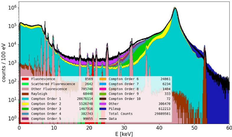
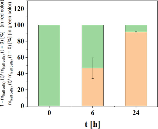
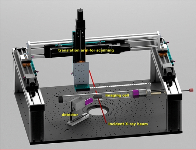
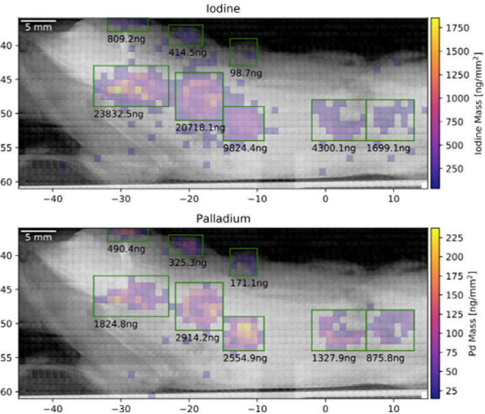
The infiltration of immune cells into sites of inflammation is one key feature of immune mediated inflammatory diseases. A detailed assessment of the in vivo dynamics of relevant cell subtypes could booster the understanding of this disease and the development of novel therapies. We show in detail how advanced X-ray fluorescence imaging enables such quantitative in vivo cell tracking, offering solutions that could pave the way beyond what other imaging modalities provide today. The key for this achievement is a detailed study of the spectral background contribution from multiple Compton scattering in a mouse-scaled object when this is scanned with a monochromatic pencil X-ray beam from a synchrotron. Under optimal conditions, the detection sensitivity is sufficient for detecting local accumulations of the labelled immune cells, hence providing experimental demonstration of in vivo immune cell tracking in mice.

摘要

免疫细胞浸润到炎症部位是免疫介导的炎症性疾病的一个关键特征。对相关细胞亚型的体内动力学进行详细评估,可以增进对这种疾病的理解,并开发新的治疗方法。我们详细展示了先进的 X 射线荧光成像如何实现这种定量的体内细胞跟踪,提供的解决方案可能超越其他成像模式目前所能提供的。这一成就的关键是在对小鼠大小的物体进行同步加速器单色铅笔 X 射线扫描时,对多次康普顿散射的光谱背景贡献进行详细研究。在最佳条件下,检测灵敏度足以检测到标记免疫细胞的局部积聚,从而在小鼠体内进行了免疫细胞跟踪的实验演示。

https://cdn.ncbi.nlm.nih.gov/pmc/blobs/e3aa/10352369/e8feabbabb89/41598_2023_38536_Fig11_HTML.jpg
https://cdn.ncbi.nlm.nih.gov/pmc/blobs/e3aa/10352369/4ea462e01df4/41598_2023_38536_Fig1_HTML.jpg
https://cdn.ncbi.nlm.nih.gov/pmc/blobs/e3aa/10352369/a0c0e5ce9cb9/41598_2023_38536_Fig2_HTML.jpg
https://cdn.ncbi.nlm.nih.gov/pmc/blobs/e3aa/10352369/616f98b00f20/41598_2023_38536_Fig3_HTML.jpg
https://cdn.ncbi.nlm.nih.gov/pmc/blobs/e3aa/10352369/ebe8e71f062a/41598_2023_38536_Fig4_HTML.jpg
https://cdn.ncbi.nlm.nih.gov/pmc/blobs/e3aa/10352369/6f1e624e59ad/41598_2023_38536_Fig5_HTML.jpg
https://cdn.ncbi.nlm.nih.gov/pmc/blobs/e3aa/10352369/085ef47c035f/41598_2023_38536_Fig6_HTML.jpg
https://cdn.ncbi.nlm.nih.gov/pmc/blobs/e3aa/10352369/9f7763dfb54d/41598_2023_38536_Fig7_HTML.jpg
https://cdn.ncbi.nlm.nih.gov/pmc/blobs/e3aa/10352369/8795051a914c/41598_2023_38536_Fig8_HTML.jpg
https://cdn.ncbi.nlm.nih.gov/pmc/blobs/e3aa/10352369/332ba0b408ac/41598_2023_38536_Fig9_HTML.jpg
https://cdn.ncbi.nlm.nih.gov/pmc/blobs/e3aa/10352369/513e7e21670b/41598_2023_38536_Fig10_HTML.jpg
https://cdn.ncbi.nlm.nih.gov/pmc/blobs/e3aa/10352369/e8feabbabb89/41598_2023_38536_Fig11_HTML.jpg

相似文献

1
Enabling X-ray fluorescence imaging for in vivo immune cell tracking.实现体内免疫细胞追踪的 X 射线荧光成像。
Sci Rep. 2023 Jul 17;13(1):11505. doi: 10.1038/s41598-023-38536-5.
2
First Demonstration of Compton Camera Used for X-Ray Fluorescence Imaging.首次展示用于 X 射线荧光成像的康普顿相机。
IEEE Trans Med Imaging. 2023 May;42(5):1314-1324. doi: 10.1109/TMI.2022.3226329. Epub 2023 May 2.
3
X-ray fluorescence computed tomography (XFCT) imaging of gold nanoparticle-loaded objects using 110 kVp x-rays.采用 110kVp X 射线对负载金纳米粒子的物体进行 X 射线荧光计算机断层成像(XFCT)。
Phys Med Biol. 2010 Feb 7;55(3):647-62. doi: 10.1088/0031-9155/55/3/007. Epub 2010 Jan 13.
4
Feasibility of Monitoring Tumor Response by Tracking Nanoparticle-Labelled T Cells Using X-ray Fluorescence Imaging-A Numerical Study.基于 X 射线荧光成像监测标记纳米颗粒 T 细胞以评估肿瘤应答的可行性:数值研究。
Int J Mol Sci. 2021 Aug 14;22(16):8736. doi: 10.3390/ijms22168736.
5
Review of Development and Recent Advances in Biomedical X-ray Fluorescence Imaging.生物医学 X 射线荧光成像的发展与最新进展综述。
Int J Mol Sci. 2023 Jul 1;24(13):10990. doi: 10.3390/ijms241310990.
6
Broadband, monochromatic and quasi-monochromatic x-ray propagation in multi-Z media for imaging and diagnostics.用于成像和诊断的多Z介质中的宽带、单色和准单色X射线传播
Phys Med Biol. 2017 Jul 20;62(16):6361-6378. doi: 10.1088/1361-6560/aa7cd6.
7
Feasibility study of Compton cameras for x-ray fluorescence computed tomography with humans.用于人体X射线荧光计算机断层扫描的康普顿相机可行性研究。
Phys Med Biol. 2016 Dec 21;61(24):8521-8540. doi: 10.1088/0031-9155/61/24/8521. Epub 2016 Nov 15.
8
Enabling Coarse X-ray Fluorescence Imaging Scans with Enlarged Synchrotron Beam by Means of Mosaic Crystal Defocusing Optics.利用镶嵌晶体离焦光学实现同步加速器光束放大的粗 X 射线荧光成像扫描。
Int J Mol Sci. 2022 Apr 23;23(9):4673. doi: 10.3390/ijms23094673.
9
X-ray phase-contrast tomography with a compact laser-driven synchrotron source.采用紧凑型激光驱动同步加速器源的X射线相衬断层扫描术。
Proc Natl Acad Sci U S A. 2015 May 5;112(18):5567-72. doi: 10.1073/pnas.1500938112. Epub 2015 Apr 20.
10
Spectroscopic imaging at compact inverse Compton X-ray sources.紧凑型逆康普顿 X 射线源的光谱成象。
Phys Med. 2020 Nov;79:137-144. doi: 10.1016/j.ejmp.2020.11.015. Epub 2020 Nov 30.

引用本文的文献

1
Biodistribution of nanoplastics in mice: advancing analytical techniques using metal-doped plastics.纳米塑料在小鼠体内的生物分布:利用金属掺杂塑料改进分析技术
Commun Biol. 2025 Aug 19;8(1):1247. doi: 10.1038/s42003-025-08709-1.
2
Voices of Nanomedicine: Blueprint Guidelines for Collaboration in Addressing Global Unmet Medical Needs.纳米医学之声:应对全球未满足医疗需求的合作蓝图指南
ACS Nano. 2025 Jan 28;19(3):2979-2991. doi: 10.1021/acsnano.4c13513. Epub 2025 Jan 10.
3
Numerical Study towards In Vivo Tracking of Micro-/Nanoplastic Based on X-ray Fluorescence Imaging.

本文引用的文献

1
Compact all-optical precision-tunable narrowband hard Compton X-ray source.紧凑型全光精密可调谐窄带硬康普顿X射线源。
Sci Rep. 2022 Sep 26;12(1):16017. doi: 10.1038/s41598-022-20283-8.
2
Assessing Cellular Uptake of Exogenous Coenzyme Q into Human Skin Cells by X-ray Fluorescence Imaging.通过X射线荧光成像评估外源性辅酶Q进入人皮肤细胞的细胞摄取情况。
Antioxidants (Basel). 2022 Aug 6;11(8):1532. doi: 10.3390/antiox11081532.
3
Quantitative considerations about the size dependence of cellular entry and excretion of colloidal nanoparticles for different cell types.
基于X射线荧光成像的微/纳米塑料体内追踪数值研究
Biomedicines. 2024 Jul 5;12(7):1500. doi: 10.3390/biomedicines12071500.
4
Enabling Low-Dose In Vivo Benchtop X-ray Fluorescence Computed Tomography through Deep-Learning-Based Denoising.通过基于深度学习的去噪实现低剂量活体台式X射线荧光计算机断层扫描
J Imaging. 2024 May 22;10(6):127. doi: 10.3390/jimaging10060127.
5
High-Spatial-Resolution Benchtop X-ray Fluorescence Imaging through Bragg-Diffraction-Based Focusing with Bent Mosaic Graphite Crystals: A Simulation Study.基于弯曲镶嵌石墨晶体的布拉格衍射聚焦的高空间分辨率台式 X 射线荧光成像:模拟研究。
Int J Mol Sci. 2024 Apr 26;25(9):4733. doi: 10.3390/ijms25094733.
6
Laboratory Liquid-Jet X-ray Microscopy and X-ray Fluorescence Imaging for Biomedical Applications.实验室液体喷射 X 射线显微镜和 X 射线荧光成像在生物医学中的应用。
Int J Mol Sci. 2024 Jan 11;25(2):920. doi: 10.3390/ijms25020920.
关于不同细胞类型中胶体纳米颗粒的细胞摄取和排泄的尺寸依赖性的定量考量。
ChemTexts. 2022;8(1):9. doi: 10.1007/s40828-021-00159-6. Epub 2022 Jan 25.
4
In-situ x-ray fluorescence imaging of the endogenous iodine distribution in murine thyroids.在体 X 射线荧光成像检测小鼠甲状腺内源性碘分布。
Sci Rep. 2022 Feb 21;12(1):2903. doi: 10.1038/s41598-022-06786-4.
5
X-ray-Fluorescence Imaging for In Vivo Detection of Gold-Nanoparticle-Labeled Immune Cells: A GEANT4 Based Feasibility Study.用于体内检测金纳米颗粒标记免疫细胞的X射线荧光成像:基于GEANT4的可行性研究
Cancers (Basel). 2021 Nov 17;13(22):5759. doi: 10.3390/cancers13225759.
6
IL-1-driven stromal-neutrophil interactions define a subset of patients with inflammatory bowel disease that does not respond to therapies.IL-1 驱动的基质-中性粒细胞相互作用定义了一部分对治疗无反应的炎症性肠病患者亚群。
Nat Med. 2021 Nov;27(11):1970-1981. doi: 10.1038/s41591-021-01520-5. Epub 2021 Oct 21.
7
Feasibility of Monitoring Tumor Response by Tracking Nanoparticle-Labelled T Cells Using X-ray Fluorescence Imaging-A Numerical Study.基于 X 射线荧光成像监测标记纳米颗粒 T 细胞以评估肿瘤应答的可行性:数值研究。
Int J Mol Sci. 2021 Aug 14;22(16):8736. doi: 10.3390/ijms22168736.
8
Single-cell analyses of Crohn's disease tissues reveal intestinal intraepithelial T cells heterogeneity and altered subset distributions.克罗恩病组织的单细胞分析揭示了肠道上皮内T细胞的异质性及亚群分布的改变。
Nat Commun. 2021 Mar 26;12(1):1921. doi: 10.1038/s41467-021-22164-6.
9
X-ray-Based Techniques to Study the Nano-Bio Interface.基于X射线的纳米-生物界面研究技术
ACS Nano. 2021 Mar 23;15(3):3754-3807. doi: 10.1021/acsnano.0c09563. Epub 2021 Mar 2.
10
Tutorial: guidelines for the computational analysis of single-cell RNA sequencing data.教程:单细胞 RNA 测序数据分析的计算分析指南。
Nat Protoc. 2021 Jan;16(1):1-9. doi: 10.1038/s41596-020-00409-w. Epub 2020 Dec 7.