Majrashi Majed, Kotowska Anna, Scurr David, Hicks Jacqueline M, Ghaemmaghami Amir, Yang Jing
School of Pharmacy, University of Nottingham, Nottingham NG7 2RD, U.K.
Biodiscovery Institute, University of Nottingham, Nottingham NG7 2RD, U.K.
ACS Appl Mater Interfaces. 2023 Nov 28;15(49):56623-38. doi: 10.1021/acsami.3c09774.
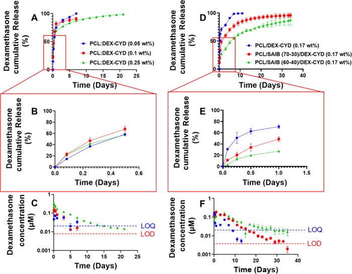
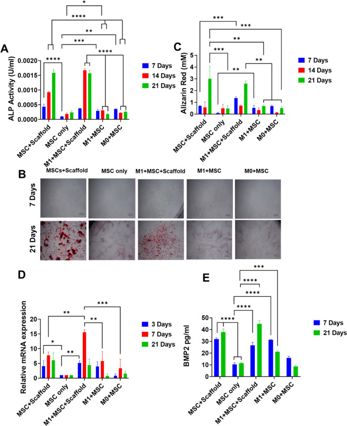
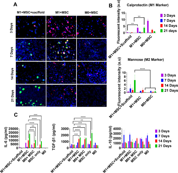
Enhancing osteogenesis via modulating immune cells is emerging as a new approach to address the current challenges in repairing bone defects and fractures. However, much remains unknown about the crosstalk between immune cells and osteolineage cells during bone formation. Moreover, biomaterial scaffold-based approaches to effectively modulate this crosstalk to favor bone healing are also lacking. This study is the first to investigate the interactions between macrophages and mesenchymal stem cells (MSCs) in co-cultures with the sustained release of an anti-inflammatory and pro-osteogenesis drug (dexamethasone) from three-dimensional (3D)-printed scaffolds. We successfully achieved the sustained release of dexamethasone from polycaprolactone (PCL) by adding the excipient-sucrose acetate isobutyrate (SAIB). Dexamethasone was released over 35 days in the 17-163 nM range. The osteogenic differentiation of MSCs was enhanced by M1 macrophages at early time points. The late-stage mineralization was dominated by dexamethasone, with little contribution from the macrophages. Besides confirming BMP-2 whose secretion was promoted by both dexamethasone and M1 macrophages as a soluble mediator for enhanced osteogenesis, IL-6 was found to be a possible new soluble factor that mediated osteogenesis in macrophage-MSC co-cultures. The phenotype switching from M1 to M2 was drastically enhanced by the scaffold-released dexamethasone but only marginally by the co-cultured MSCs. Our results offer new insight into macrophage-MSC crosstalk and demonstrate the potential of using drug-release scaffolds to both modulate inflammation and enhance bone regeneration.
通过调节免疫细胞来增强骨生成正成为应对当前骨缺损和骨折修复挑战的一种新方法。然而,在骨形成过程中免疫细胞与骨谱系细胞之间的相互作用仍有许多未知之处。此外,基于生物材料支架有效调节这种相互作用以促进骨愈合的方法也很缺乏。本研究首次探究了巨噬细胞与间充质干细胞(MSCs)在共培养中的相互作用,其中抗炎和促骨生成药物(地塞米松)从三维(3D)打印支架中持续释放。我们通过添加辅料醋酸异丁酸蔗糖酯(SAIB)成功实现了地塞米松从聚己内酯(PCL)中的持续释放。地塞米松在35天内以17 - 163 nM的浓度范围释放。在早期阶段,M1巨噬细胞增强了MSCs的成骨分化。后期矿化主要由地塞米松主导,巨噬细胞的贡献很小。除了证实地塞米松和M1巨噬细胞均促进其分泌的BMP - 2作为增强成骨的可溶性介质外,还发现IL - 6是巨噬细胞 - MSC共培养中介导成骨的一种可能的新可溶性因子。支架释放的地塞米松极大地增强了从M1到M2的表型转换,但共培养的MSCs仅略微增强了这种转换。我们的结果为巨噬细胞 - MSC相互作用提供了新的见解,并证明了使用药物释放支架调节炎症和增强骨再生的潜力。