• 文献检索
  • 文档翻译
  • 深度研究
  • 学术资讯
  • Suppr Zotero 插件Zotero 插件
  • 邀请有礼
  • 套餐&价格
  • 历史记录
应用&插件
Suppr Zotero 插件Zotero 插件浏览器插件Mac 客户端Windows 客户端微信小程序
定价
高级版会员购买积分包购买API积分包
服务
文献检索文档翻译深度研究API 文档MCP 服务
关于我们
关于 Suppr公司介绍联系我们用户协议隐私条款
关注我们

Suppr 超能文献

核心技术专利:CN118964589B侵权必究
粤ICP备2023148730 号-1Suppr @ 2026

文献检索

告别复杂PubMed语法,用中文像聊天一样搜索,搜遍4000万医学文献。AI智能推荐,让科研检索更轻松。

立即免费搜索

文件翻译

保留排版,准确专业,支持PDF/Word/PPT等文件格式,支持 12+语言互译。

免费翻译文档

深度研究

AI帮你快速写综述,25分钟生成高质量综述,智能提取关键信息,辅助科研写作。

立即免费体验

血细胞图像分割与分类:系统综述

Blood cell image segmentation and classification: a systematic review.

作者信息

Shahzad Muhammad, Ali Farman, Shirazi Syed Hamad, Rasheed Assad, Ahmad Awais, Shah Babar, Kwak Daehan

机构信息

Department of Computer Science and Information Technology, Hazara University, Mansehra, Pakistan.

Department of Computer Science and Engineering, School of Convergence, Sungkyunkwan University, Seoul, South Korea.

出版信息

PeerJ Comput Sci. 2024 Feb 2;10:e1813. doi: 10.7717/peerj-cs.1813. eCollection 2024.

DOI:10.7717/peerj-cs.1813
PMID:38435563
原文链接:https://pmc.ncbi.nlm.nih.gov/articles/PMC10909159/
Abstract

BACKGROUND

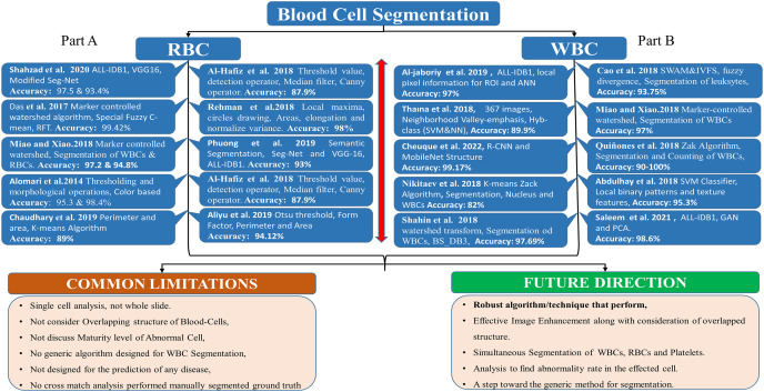
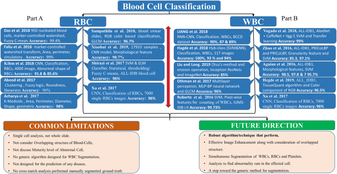
Blood diseases such as leukemia, anemia, lymphoma, and thalassemia are hematological disorders that relate to abnormalities in the morphology and concentration of blood elements, specifically white blood cells (WBC) and red blood cells (RBC). Accurate and efficient diagnosis of these conditions significantly depends on the expertise of hematologists and pathologists. To assist the pathologist in the diagnostic process, there has been growing interest in utilizing computer-aided diagnostic (CAD) techniques, particularly those using medical image processing and machine learning algorithms. Previous surveys in this domain have been narrowly focused, often only addressing specific areas like segmentation or classification but lacking a holistic view like segmentation, classification, feature extraction, dataset utilization, evaluation matrices, .

METHODOLOGY

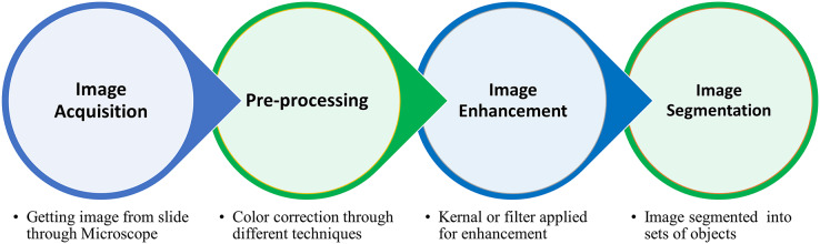
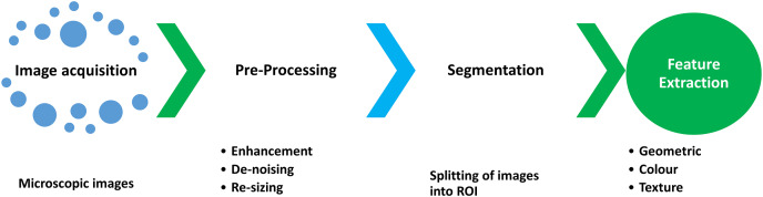
This survey aims to provide a comprehensive and systematic review of existing literature and research work in the field of blood image analysis using deep learning techniques. It particularly focuses on medical image processing techniques and deep learning algorithms that excel in the morphological characterization of WBCs and RBCs. The review is structured to cover four main areas: segmentation techniques, classification methodologies, descriptive feature selection, evaluation parameters, and dataset selection for the analysis of WBCs and RBCs.

RESULTS

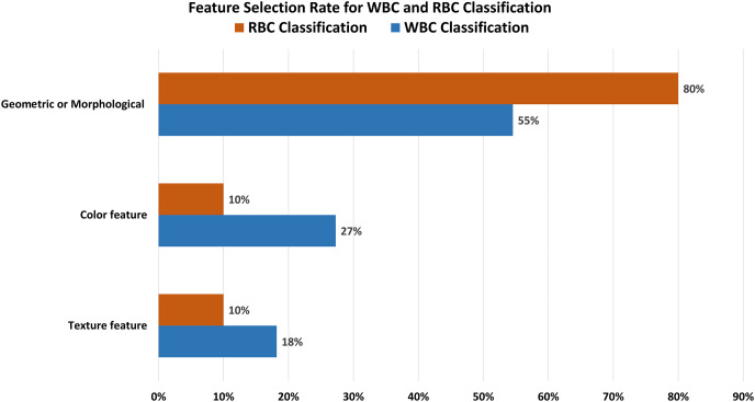
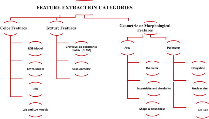
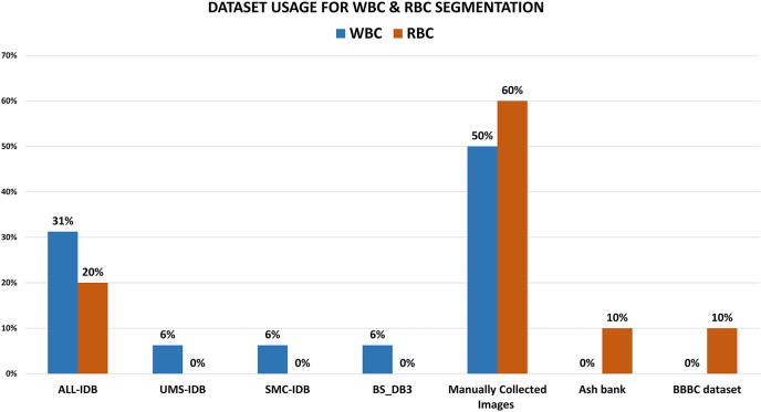
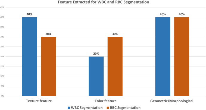
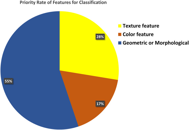
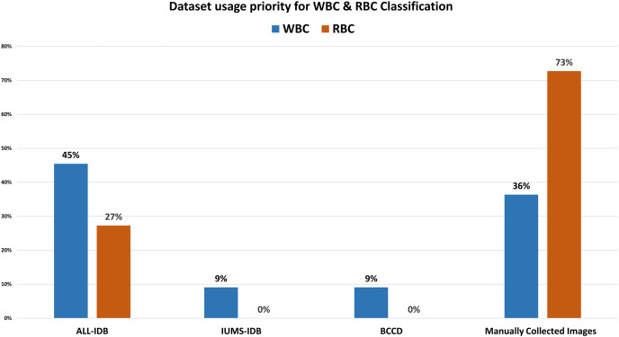
Our analysis reveals several interesting trends and preferences among researchers. Regarding dataset selection, approximately 50% of research related to WBC segmentation and 60% for RBC segmentation opted for manually obtaining images rather than using a predefined dataset. When it comes to classification, 45% of the previous work on WBCs chose the ALL-IDB dataset, while a significant 73% of researchers focused on RBC classification decided to manually obtain images from medical institutions instead of utilizing predefined datasets. In terms of feature selection for classification, morphological features were the most popular, being chosen in 55% and 80% of studies related to WBC and RBC classification, respectively.

CONCLUSION

The diagnostic accuracy for blood-related diseases like leukemia, anemia, lymphoma, and thalassemia can be significantly enhanced through the effective use of CAD techniques, which have evolved considerably in recent years. This survey provides a broad and in-depth review of the techniques being employed, from image segmentation to classification, feature selection, utilization of evaluation matrices, and dataset selection. The inconsistency in dataset selection suggests a need for standardized, high-quality datasets to strengthen the diagnostic capabilities of these techniques further. Additionally, the popularity of morphological features indicates that future research could further explore and innovate in this direction.

摘要

背景

白血病、贫血、淋巴瘤和地中海贫血等血液疾病是血液系统疾病,与血液成分,特别是白细胞(WBC)和红细胞(RBC)的形态和浓度异常有关。这些疾病的准确有效诊断很大程度上依赖于血液学家和病理学家的专业知识。为了在诊断过程中协助病理学家,人们越来越关注利用计算机辅助诊断(CAD)技术,特别是那些使用医学图像处理和机器学习算法的技术。该领域以前的调查范围较窄,通常只涉及分割或分类等特定领域,而缺乏像分割、分类、特征提取、数据集利用、评估矩阵这样的整体视角。

方法

本次调查旨在对使用深度学习技术的血液图像分析领域的现有文献和研究工作进行全面系统的综述。它特别关注在白细胞和红细胞形态特征表征方面表现出色的医学图像处理技术和深度学习算法。综述结构涵盖四个主要领域:白细胞和红细胞分析的分割技术、分类方法、描述性特征选择、评估参数和数据集选择。

结果

我们的分析揭示了研究人员之间的几个有趣趋势和偏好。在数据集选择方面,大约50%与白细胞分割相关的研究和60%与红细胞分割相关的研究选择手动获取图像,而不是使用预定义数据集。在分类方面,之前关于白细胞的工作中有45%选择了ALL-IDB数据集,而在专注于红细胞分类的研究人员中,有73%的人决定从医疗机构手动获取图像,而不是使用预定义数据集。在分类的特征选择方面,形态特征最受欢迎,在与白细胞和红细胞分类相关的研究中,分别有55%和80%的研究选择了形态特征。

结论

通过有效使用近年来有了很大发展的CAD技术,可以显著提高白血病、贫血、淋巴瘤和地中海贫血等血液相关疾病的诊断准确性。本次调查对从图像分割到分类、特征选择、评估矩阵利用和数据集选择等所采用的技术进行了广泛而深入的综述。数据集选择的不一致表明需要标准化的高质量数据集,以进一步加强这些技术的诊断能力。此外,形态特征的受欢迎程度表明未来的研究可以在这个方向上进一步探索和创新。

https://cdn.ncbi.nlm.nih.gov/pmc/blobs/a545/10909159/70d6c33bf1b5/peerj-cs-10-1813-g010.jpg
https://cdn.ncbi.nlm.nih.gov/pmc/blobs/a545/10909159/551dee55f984/peerj-cs-10-1813-g001.jpg
https://cdn.ncbi.nlm.nih.gov/pmc/blobs/a545/10909159/1c0c3e2b5d19/peerj-cs-10-1813-g002.jpg
https://cdn.ncbi.nlm.nih.gov/pmc/blobs/a545/10909159/ca224e7444bc/peerj-cs-10-1813-g003.jpg
https://cdn.ncbi.nlm.nih.gov/pmc/blobs/a545/10909159/b8ad81765b59/peerj-cs-10-1813-g004.jpg
https://cdn.ncbi.nlm.nih.gov/pmc/blobs/a545/10909159/11c44536afba/peerj-cs-10-1813-g005.jpg
https://cdn.ncbi.nlm.nih.gov/pmc/blobs/a545/10909159/90cd2ac159f5/peerj-cs-10-1813-g006.jpg
https://cdn.ncbi.nlm.nih.gov/pmc/blobs/a545/10909159/aa29e8186315/peerj-cs-10-1813-g007.jpg
https://cdn.ncbi.nlm.nih.gov/pmc/blobs/a545/10909159/f798a7540671/peerj-cs-10-1813-g008.jpg
https://cdn.ncbi.nlm.nih.gov/pmc/blobs/a545/10909159/d46ba9f90404/peerj-cs-10-1813-g009.jpg
https://cdn.ncbi.nlm.nih.gov/pmc/blobs/a545/10909159/70d6c33bf1b5/peerj-cs-10-1813-g010.jpg

相似文献

1
Blood cell image segmentation and classification: a systematic review.血细胞图像分割与分类:系统综述
PeerJ Comput Sci. 2024 Feb 2;10:e1813. doi: 10.7717/peerj-cs.1813. eCollection 2024.
2
Deep Learning Approaches Towards Skin Lesion Segmentation and Classification from Dermoscopic Images - A Review.深度学习方法在皮肤镜图像的皮肤损伤分割和分类中的应用综述。
Curr Med Imaging. 2020;16(5):513-533. doi: 10.2174/1573405615666190129120449.
3
WBC-based segmentation and classification on microscopic images: a minor improvement.基于白细胞的显微镜图像分割与分类:略有改进。
F1000Res. 2021 Nov 17;10:1168. doi: 10.12688/f1000research.73315.1. eCollection 2021.
4
GFNB: Gini index-based Fuzzy Naive Bayes and blast cell segmentation for leukemia detection using multi-cell blood smear images.基于基尼指数的模糊朴素贝叶斯和 blast 细胞分割在多细胞血涂片图像白血病检测中的应用。
Med Biol Eng Comput. 2020 Nov;58(11):2789-2803. doi: 10.1007/s11517-020-02249-y. Epub 2020 Sep 15.
5
Folic acid supplementation and malaria susceptibility and severity among people taking antifolate antimalarial drugs in endemic areas.在流行地区,服用抗叶酸抗疟药物的人群中,叶酸补充剂与疟疾易感性和严重程度的关系。
Cochrane Database Syst Rev. 2022 Feb 1;2(2022):CD014217. doi: 10.1002/14651858.CD014217.
6
Segmentation of white blood cells and comparison of cell morphology by linear and naïve Bayes classifiers.白细胞的分割以及通过线性和朴素贝叶斯分类器对细胞形态的比较。
Biomed Eng Online. 2015 Jun 30;14:63. doi: 10.1186/s12938-015-0037-1.
7
A Methodology for Texture Feature-based Quality Assessment in Nucleus Segmentation of Histopathology Image.一种基于纹理特征的组织病理学图像细胞核分割质量评估方法。
J Pathol Inform. 2017 Sep 7;8:38. doi: 10.4103/jpi.jpi_43_17. eCollection 2017.
8
Classification of white blood cells (leucocytes) from blood smear imagery using machine and deep learning models: A global scoping review.使用机器学习和深度学习模型对血涂片图像中的白细胞(白血细胞)进行分类:全球范围的综述。
PLoS One. 2024 Jun 17;19(6):e0292026. doi: 10.1371/journal.pone.0292026. eCollection 2024.
9
Nucleus segmentation of white blood cells in blood smear images by modeling the pixels' intensities as a set of three Gaussian distributions.通过将像素的强度建模为三个高斯分布的集合来对血涂片图像中的白细胞进行核分割。
Med Biol Eng Comput. 2024 Aug;62(8):2371-2388. doi: 10.1007/s11517-024-03065-4. Epub 2024 Apr 8.
10
White blood cells detection and classification based on regional convolutional neural networks.基于区域卷积神经网络的白细胞检测与分类。
Med Hypotheses. 2020 Feb;135:109472. doi: 10.1016/j.mehy.2019.109472. Epub 2019 Nov 4.

引用本文的文献

1
Enhanced detection of malaria infected red blood cells through phase driven classification.通过相位驱动分类增强对疟疾感染红细胞的检测。
Sci Rep. 2025 Aug 21;15(1):30733. doi: 10.1038/s41598-025-12899-3.
2
Advancements in Circulating Tumor Cell Detection for Early Cancer Diagnosis: An Integration of Machine Learning Algorithms with Microfluidic Technologies.用于早期癌症诊断的循环肿瘤细胞检测进展:机器学习算法与微流控技术的整合
Biosensors (Basel). 2025 Mar 29;15(4):220. doi: 10.3390/bios15040220.
3
Revolutionizing diagnosis of pulmonary based on CT: a systematic review of imaging analysis through deep learning.

本文引用的文献

1
Classification of white blood cells (leucocytes) from blood smear imagery using machine and deep learning models: A global scoping review.使用机器学习和深度学习模型对血涂片图像中的白细胞(白血细胞)进行分类:全球范围的综述。
PLoS One. 2024 Jun 17;19(6):e0292026. doi: 10.1371/journal.pone.0292026. eCollection 2024.
2
A deep learning based dual encoder-decoder framework for anatomical structure segmentation in chest X-ray images.基于深度学习的双编码器-解码器框架,用于胸部 X 光图像中的解剖结构分割。
Sci Rep. 2023 Jan 16;13(1):791. doi: 10.1038/s41598-023-27815-w.
3
WBC image classification and generative models based on convolutional neural network.
基于CT的肺部诊断革命:深度学习影像分析的系统综述
Front Microbiol. 2025 Jan 8;15:1510026. doi: 10.3389/fmicb.2024.1510026. eCollection 2024.
基于卷积神经网络的白细胞图像分类与生成模型。
BMC Med Imaging. 2022 May 20;22(1):94. doi: 10.1186/s12880-022-00818-1.
4
An Efficient Multi-Level Convolutional Neural Network Approach for White Blood Cells Classification.一种用于白细胞分类的高效多级卷积神经网络方法。
Diagnostics (Basel). 2022 Jan 20;12(2):248. doi: 10.3390/diagnostics12020248.
5
A large dataset of white blood cells containing cell locations and types, along with segmented nuclei and cytoplasm.一个包含细胞位置和类型、以及分割后的细胞核和细胞质的白细胞大型数据集。
Sci Rep. 2022 Jan 21;12(1):1123. doi: 10.1038/s41598-021-04426-x.
6
Robust Method for Semantic Segmentation of Whole-Slide Blood Cell Microscopic Images.全玻片血细胞质子显微镜图像语义分割的稳健方法。
Comput Math Methods Med. 2020 Jan 21;2020:4015323. doi: 10.1155/2020/4015323. eCollection 2020.
7
Evaluation of Deep Learning Strategies for Nucleus Segmentation in Fluorescence Images.深度学习策略在荧光图像中细胞核分割的评估。
Cytometry A. 2019 Sep;95(9):952-965. doi: 10.1002/cyto.a.23863. Epub 2019 Jul 16.
8
Automatic classification of cells in microscopic fecal images using convolutional neural networks.使用卷积神经网络对显微镜下粪便图像中的细胞进行自动分类。
Biosci Rep. 2019 Apr 5;39(4). doi: 10.1042/BSR20182100. Print 2019 Apr 30.
9
Acute Lymphoblastic Leukemia Detection and Classification of Its Subtypes Using Pretrained Deep Convolutional Neural Networks.使用预训练深度卷积神经网络检测急性淋巴细胞白血病及其亚型分类
Technol Cancer Res Treat. 2018 Jan 1;17:1533033818802789. doi: 10.1177/1533033818802789.
10
Classification of red blood cell shapes in flow using outlier tolerant machine learning.使用容忍异常值的机器学习对红细胞形状进行分类。
PLoS Comput Biol. 2018 Jun 15;14(6):e1006278. doi: 10.1371/journal.pcbi.1006278. eCollection 2018 Jun.