School of Materials Science and Engineering, Hebei University of Technology, Tianjin, People's Republic of China.
Center for Health Science and Engineering, Hebei Key Laboratory of Biomaterials and Smart Theranostics, School of Health Sciences and Biomedical Engineering, Hebei University of Technology, Tianjin, 300131, People's Republic of China.
Int J Nanomedicine. 2024 Jun 1;19:5109-5123. doi: 10.2147/IJN.S460339. eCollection 2024.
Lumbar interbody fusion is widely employed for both acute and chronic spinal diseases interventions. However, large incision created during interbody cage implantation may adversely impair spinal tissue and influence postoperative recovery. The aim of this study was to design a shape memory interbody fusion device suitable for small incision implantation.
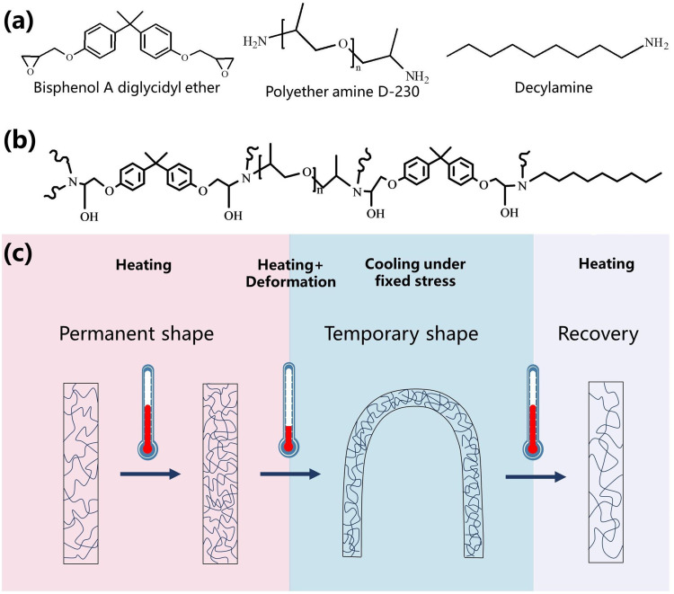
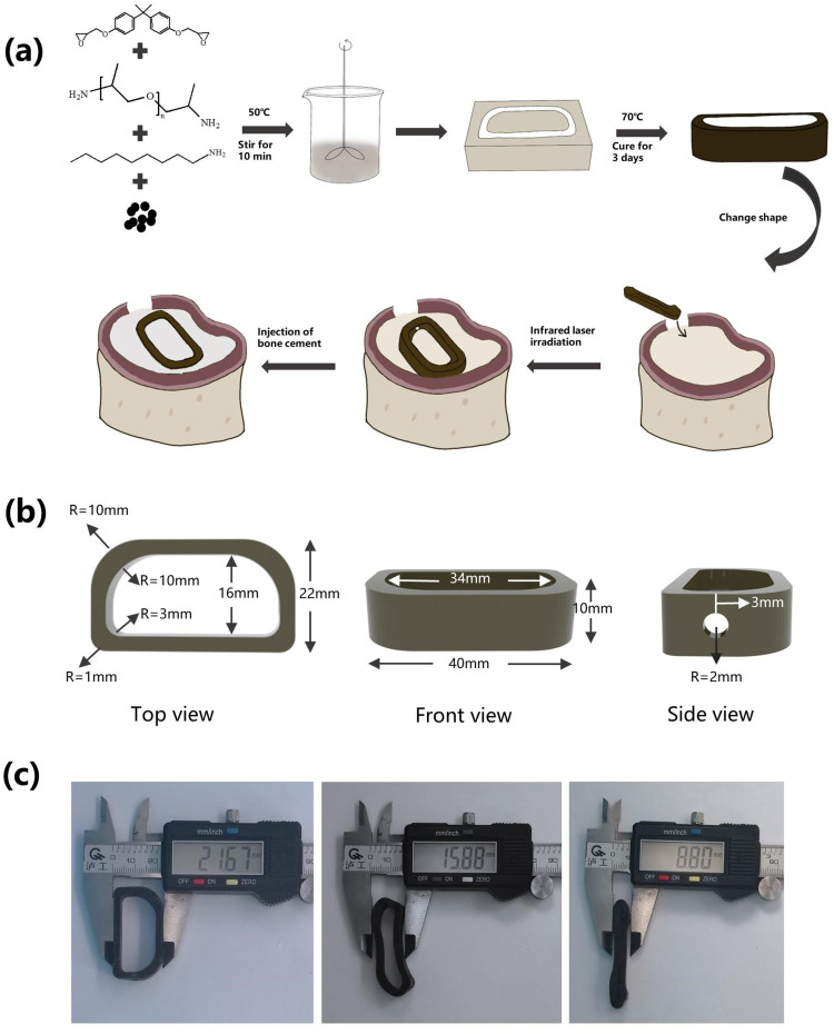
In this study, we designed and fabricated an intervertebral fusion cage that utilizes near-infrared (NIR) light-responsive shape memory characteristics. This cage was composed of bisphenol A diglycidyl ether, polyether amine D-230, decylamine and iron oxide nanoparticles. A self-hardening calcium phosphate-starch cement (CSC) was injected internally through the injection channel of the cage for healing outcome improvement.
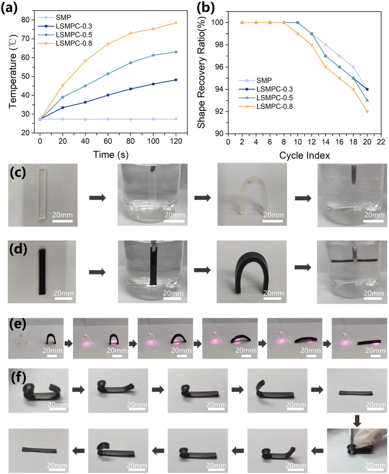
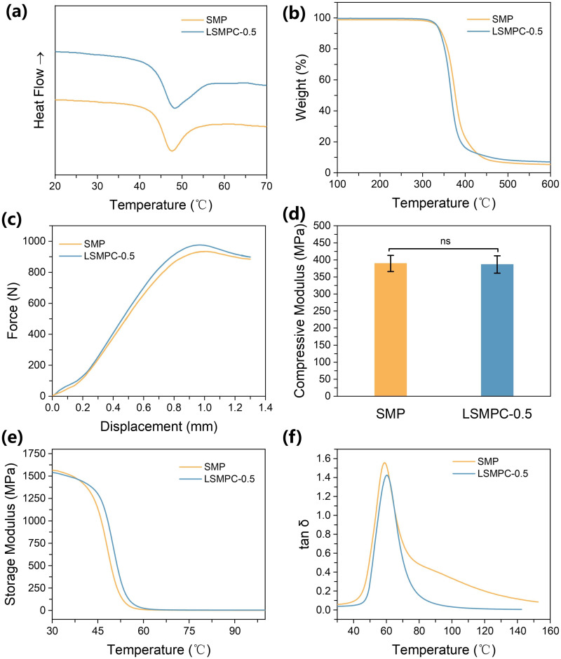
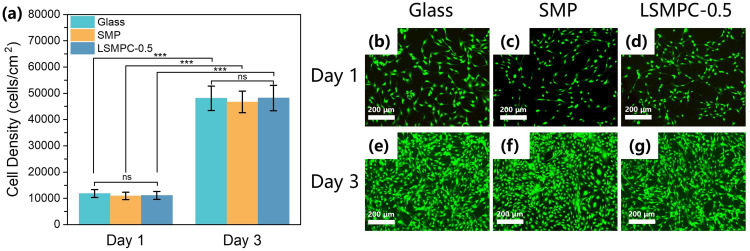
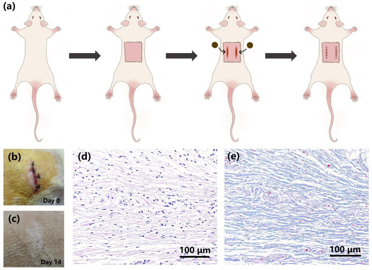
The size of the interbody cage is reduced from 22 mm to 8.8 mm to minimize the incision size. Subsequent NIR light irradiation prompted a swift recovery of the cage shape within 5 min at the lesion site. The biocompatibility of the shape memory composite was validated through in vitro MC3T3-E1 cell (osteoblast-like cells) adhesion and proliferation assays and subcutaneous implantation experiments in rats. CSC was injected into the cage, and the relevant results revealed that CSC is uniformly dispersed within the internal space, along with the cage compressive strength increasing from 12 to 20 MPa.
The results from this study thus demonstrated that this integrated approach of using a minimally invasive NIR shape memory spinal fusion cage with CSC has potential for lumbar interbody fusion.
腰椎体间融合术广泛应用于急性和慢性脊柱疾病的干预治疗。然而,在椎间笼植入过程中产生的大切口可能会对脊柱组织造成不良影响,并影响术后恢复。本研究旨在设计一种适用于小切口植入的形状记忆体间融合装置。
本研究设计并制造了一种利用近红外(NIR)光响应形状记忆特性的椎间融合笼。该笼由双酚 A 二缩水甘油醚、聚醚胺 D-230、癸胺和氧化铁纳米粒子组成。通过笼的注射通道内部注入自硬磷酸钙-淀粉水泥(CSC),以改善愈合效果。
椎间笼的尺寸从 22mm 减小到 8.8mm,以最小化切口尺寸。随后的 NIR 光照射在病变部位 5 分钟内迅速恢复了笼的形状。通过体外 MC3T3-E1 细胞(成骨样细胞)黏附和增殖实验以及大鼠皮下植入实验验证了形状记忆复合材料的生物相容性。CSC 被注入笼内,相关结果表明 CSC 均匀分散在内部空间内,同时笼的压缩强度从 12MPa 增加到 20MPa。
因此,本研究结果表明,使用具有 CSC 的微创 NIR 形状记忆脊柱融合笼的综合方法具有腰椎体间融合的潜力。