Department of Biochemistry, Schulich School of Medicine & Dentistry, Western University, London, Ontario, Canada.
Department of Computer Science, Western University, London, Ontario, Canada.
PLoS Comput Biol. 2024 Jun 27;20(6):e1012254. doi: 10.1371/journal.pcbi.1012254. eCollection 2024 Jun.
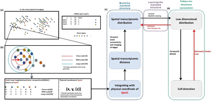
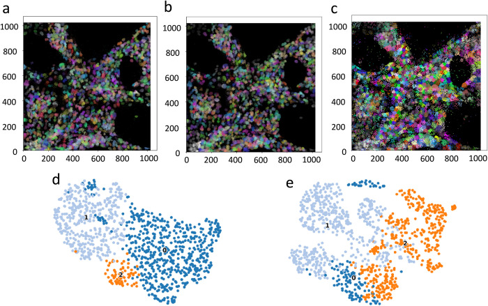
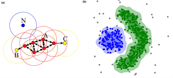
Spatial transcriptomics has gained popularity over the past decade due to its ability to evaluate transcriptome data while preserving spatial information. Cell segmentation is a crucial step in spatial transcriptomic analysis, as it enables the avoidance of unpredictable tissue disentanglement steps. Although high-quality cell segmentation algorithms can aid in the extraction of valuable data, traditional methods are frequently non-spatial, do not account for spatial information efficiently, and perform poorly when confronted with the problem of spatial transcriptome cell segmentation with varying shapes. In this study, we propose ST-CellSeg, an image-based machine learning method for spatial transcriptomics that uses manifold for cell segmentation and is novel in its consideration of multi-scale information. We first construct a fully connected graph which acts as a spatial transcriptomic manifold. Using multi-scale data, we then determine the low-dimensional spatial probability distribution representation for cell segmentation. Using the adjusted Rand index (ARI), normalized mutual information (NMI), and Silhouette coefficient (SC) as model performance measures, the proposed algorithm significantly outperforms baseline models in selected datasets and is efficient in computational complexity.
空间转录组学在过去十年中变得流行起来,因为它能够在保留空间信息的同时评估转录组数据。细胞分割是空间转录组分析的关键步骤,因为它可以避免不可预测的组织分离步骤。虽然高质量的细胞分割算法可以帮助提取有价值的数据,但传统方法通常是非空间的,不能有效地利用空间信息,并且在面对具有不同形状的空间转录组细胞分割问题时表现不佳。在这项研究中,我们提出了 ST-CellSeg,这是一种基于图像的机器学习方法,用于空间转录组学,它使用流形进行细胞分割,并考虑了多尺度信息。我们首先构建了一个全连接图,作为空间转录组学的流形。然后,我们使用多尺度数据确定细胞分割的低维空间概率分布表示。使用调整后的兰德指数(ARI)、归一化互信息(NMI)和轮廓系数(SC)作为模型性能指标,所提出的算法在选定的数据集上显著优于基线模型,并且在计算复杂度方面效率很高。