• 文献检索
  • 文档翻译
  • 深度研究
  • 学术资讯
  • Suppr Zotero 插件Zotero 插件
  • 邀请有礼
  • 套餐&价格
  • 历史记录
应用&插件
Suppr Zotero 插件Zotero 插件浏览器插件Mac 客户端Windows 客户端微信小程序
定价
高级版会员购买积分包购买API积分包
服务
文献检索文档翻译深度研究API 文档MCP 服务
关于我们
关于 Suppr公司介绍联系我们用户协议隐私条款
关注我们

Suppr 超能文献

核心技术专利:CN118964589B侵权必究
粤ICP备2023148730 号-1Suppr @ 2026

文献检索

告别复杂PubMed语法,用中文像聊天一样搜索,搜遍4000万医学文献。AI智能推荐,让科研检索更轻松。

立即免费搜索

文件翻译

保留排版,准确专业,支持PDF/Word/PPT等文件格式,支持 12+语言互译。

免费翻译文档

深度研究

AI帮你快速写综述,25分钟生成高质量综述,智能提取关键信息,辅助科研写作。

立即免费体验

一种使用Transformer和语言诊断脑微出血的新型检测与分类框架。

A Novel Detection and Classification Framework for Diagnosing of Cerebral Microbleeds Using Transformer and Language.

作者信息

Chen Cong, Zhao Lin-Lin, Lang Qin, Xu Yun

机构信息

School of Clinical Medicine, College of Medicine, Nanjing Medical University, Nanjing 211166, China.

Department of Neurology, The First Affiliated Hospital of Nanjing Medical University, Nanjing 210029, China.

出版信息

Bioengineering (Basel). 2024 Sep 30;11(10):993. doi: 10.3390/bioengineering11100993.

DOI:10.3390/bioengineering11100993
PMID:39451369
原文链接:https://pmc.ncbi.nlm.nih.gov/articles/PMC11504022/
Abstract

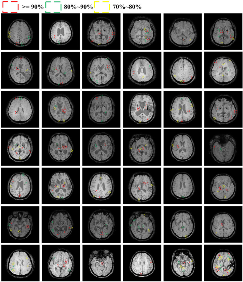
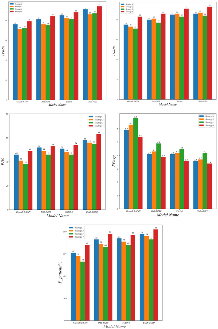
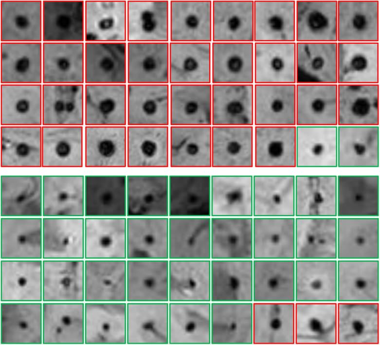
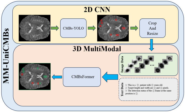
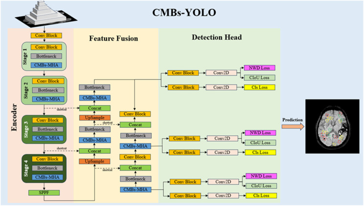
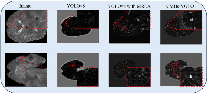
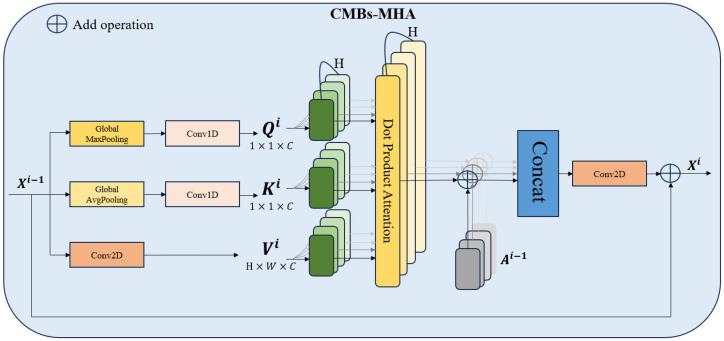
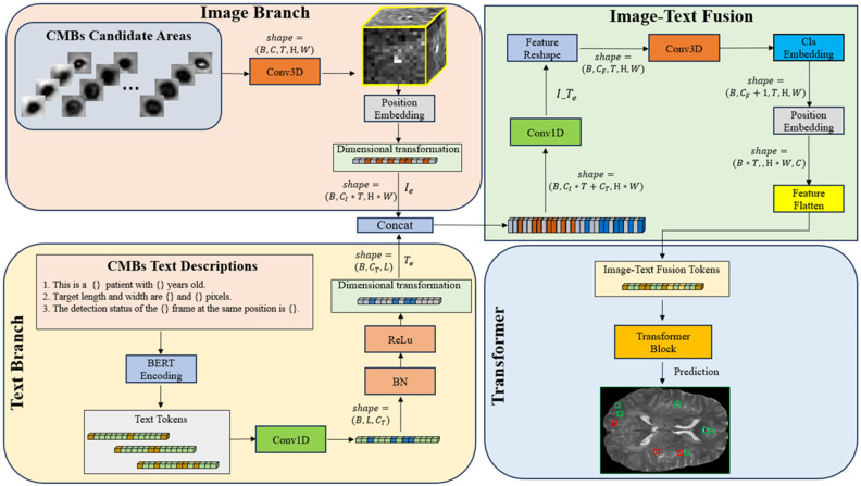
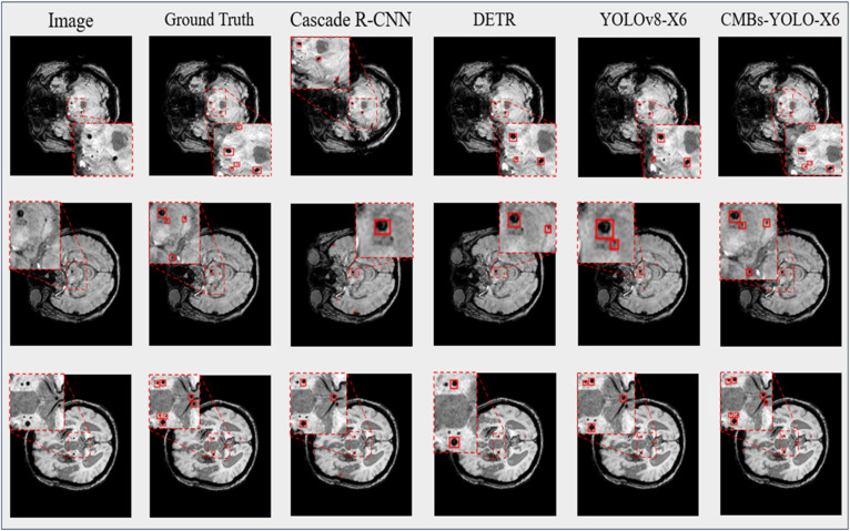
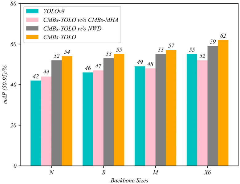
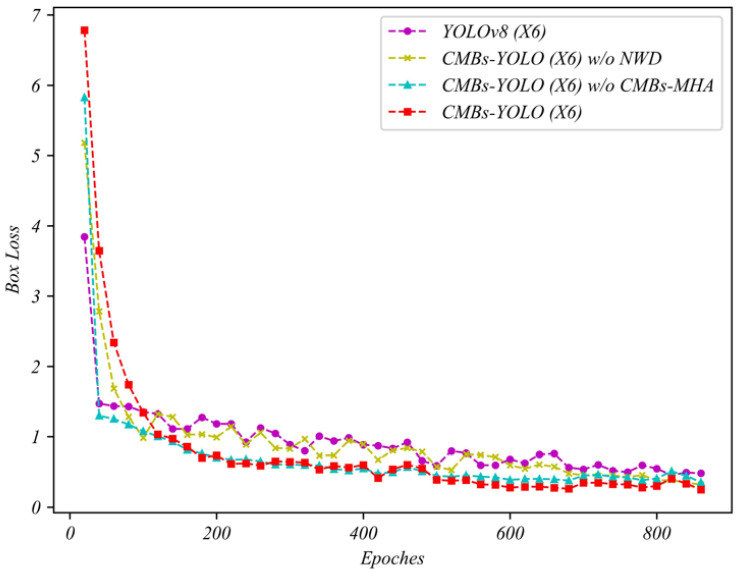
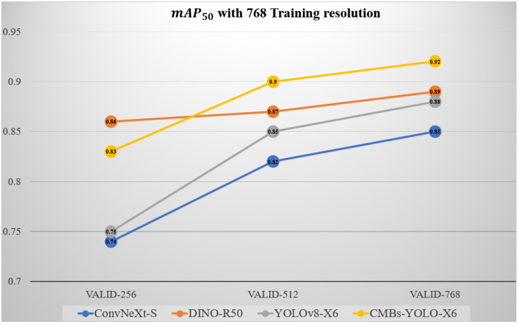
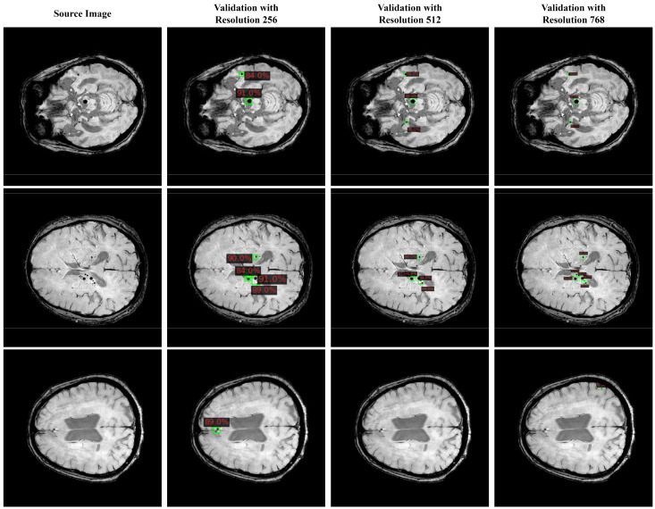
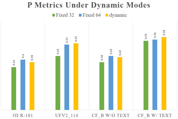
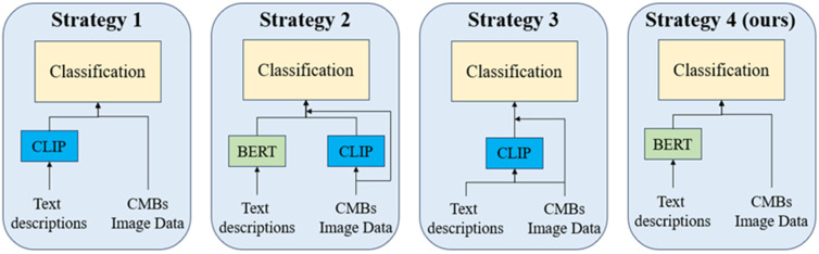
The detection of Cerebral Microbleeds (CMBs) is crucial for diagnosing cerebral small vessel disease. However, due to the small size and subtle appearance of CMBs in susceptibility-weighted imaging (SWI), manual detection is both time-consuming and labor-intensive. Meanwhile, the presence of similar-looking features in SWI images demands significant expertise from clinicians, further complicating this process. Recently, there has been a significant advancement in automated detection of CMBs using a Convolutional Neural Network (CNN) structure, aiming at enhancing diagnostic efficiency for neurologists. However, existing methods still show discrepancies when compared to the actual clinical diagnostic process. To bridge this gap, we introduce a novel multimodal detection and classification framework for CMBs' diagnosis, termed MM-UniCMBs. This framework includes a light-weight detection model and a multi-modal classification network. Specifically, we proposed a new CMBs detection network, CMBs-YOLO, designed to capture the salient features of CMBs in SWI images. Additionally, we design an innovative language-vision classification network, CMBsFormer (CF), which integrates patient textual descriptions-such as gender, age, and medical history-with image data. The MM-UniCMBs framework is designed to closely align with the diagnostic workflow of clinicians, offering greater interpretability and flexibility compared to existing methods. Extensive experimental results show that MM-UniCMBs achieves a sensitivity of 94% in CMBs' classification and can process a patient's data within 5 s.

摘要

脑微出血(CMBs)的检测对于诊断脑小血管疾病至关重要。然而,由于CMBs在磁敏感加权成像(SWI)中的尺寸小且外观不明显,手动检测既耗时又费力。同时,SWI图像中存在外观相似的特征需要临床医生具备丰富的专业知识,这使得该过程更加复杂。最近,使用卷积神经网络(CNN)结构进行CMBs自动检测取得了重大进展,旨在提高神经科医生的诊断效率。然而,与实际临床诊断过程相比,现有方法仍存在差异。为了弥合这一差距,我们引入了一种用于CMBs诊断的新型多模态检测和分类框架,称为MM-UniCMBs。该框架包括一个轻量级检测模型和一个多模态分类网络。具体而言,我们提出了一种新的CMBs检测网络CMBs-YOLO,旨在捕捉SWI图像中CMBs的显著特征。此外,我们设计了一种创新的语言-视觉分类网络CMBsFormer(CF),它将患者的文本描述(如性别、年龄和病史)与图像数据集成在一起。MM-UniCMBs框架旨在与临床医生的诊断工作流程紧密结合,与现有方法相比具有更高的可解释性和灵活性。大量实验结果表明,MM-UniCMBs在CMBs分类中实现了94%的灵敏度,并且可以在5秒内处理患者的数据。

https://cdn.ncbi.nlm.nih.gov/pmc/blobs/bc01/11504022/a702cbff28b0/bioengineering-11-00993-g020.jpg
https://cdn.ncbi.nlm.nih.gov/pmc/blobs/bc01/11504022/3c74bd7e42f5/bioengineering-11-00993-g0A1.jpg
https://cdn.ncbi.nlm.nih.gov/pmc/blobs/bc01/11504022/c8e58e5993bb/bioengineering-11-00993-g0A2.jpg
https://cdn.ncbi.nlm.nih.gov/pmc/blobs/bc01/11504022/5194864e393e/bioengineering-11-00993-g001.jpg
https://cdn.ncbi.nlm.nih.gov/pmc/blobs/bc01/11504022/274fce652b88/bioengineering-11-00993-g002.jpg
https://cdn.ncbi.nlm.nih.gov/pmc/blobs/bc01/11504022/bf340c5f86f4/bioengineering-11-00993-g003.jpg
https://cdn.ncbi.nlm.nih.gov/pmc/blobs/bc01/11504022/b76f49470027/bioengineering-11-00993-g004.jpg
https://cdn.ncbi.nlm.nih.gov/pmc/blobs/bc01/11504022/d7416e0b13e4/bioengineering-11-00993-g005.jpg
https://cdn.ncbi.nlm.nih.gov/pmc/blobs/bc01/11504022/025445bd19eb/bioengineering-11-00993-g006.jpg
https://cdn.ncbi.nlm.nih.gov/pmc/blobs/bc01/11504022/b15192e67249/bioengineering-11-00993-g007.jpg
https://cdn.ncbi.nlm.nih.gov/pmc/blobs/bc01/11504022/fb84bf1e62d1/bioengineering-11-00993-g008.jpg
https://cdn.ncbi.nlm.nih.gov/pmc/blobs/bc01/11504022/48786923f5b2/bioengineering-11-00993-g009.jpg
https://cdn.ncbi.nlm.nih.gov/pmc/blobs/bc01/11504022/83676ff85082/bioengineering-11-00993-g010.jpg
https://cdn.ncbi.nlm.nih.gov/pmc/blobs/bc01/11504022/4030c5f3cbf3/bioengineering-11-00993-g011.jpg
https://cdn.ncbi.nlm.nih.gov/pmc/blobs/bc01/11504022/afde6f796e49/bioengineering-11-00993-g012.jpg
https://cdn.ncbi.nlm.nih.gov/pmc/blobs/bc01/11504022/d746fc592528/bioengineering-11-00993-g013.jpg
https://cdn.ncbi.nlm.nih.gov/pmc/blobs/bc01/11504022/bf22baf4503a/bioengineering-11-00993-g014.jpg
https://cdn.ncbi.nlm.nih.gov/pmc/blobs/bc01/11504022/aa1eb24ff225/bioengineering-11-00993-g015.jpg
https://cdn.ncbi.nlm.nih.gov/pmc/blobs/bc01/11504022/99df868e9c28/bioengineering-11-00993-g016.jpg
https://cdn.ncbi.nlm.nih.gov/pmc/blobs/bc01/11504022/371759d3d369/bioengineering-11-00993-g017.jpg
https://cdn.ncbi.nlm.nih.gov/pmc/blobs/bc01/11504022/0fc04aa8fbd0/bioengineering-11-00993-g018.jpg
https://cdn.ncbi.nlm.nih.gov/pmc/blobs/bc01/11504022/bc047edef642/bioengineering-11-00993-g019.jpg
https://cdn.ncbi.nlm.nih.gov/pmc/blobs/bc01/11504022/a702cbff28b0/bioengineering-11-00993-g020.jpg

相似文献

1
A Novel Detection and Classification Framework for Diagnosing of Cerebral Microbleeds Using Transformer and Language.一种使用Transformer和语言诊断脑微出血的新型检测与分类框架。
Bioengineering (Basel). 2024 Sep 30;11(10):993. doi: 10.3390/bioengineering11100993.
2
Automated detection of cerebral microbleeds in MR images: A two-stage deep learning approach.基于两阶段深度学习的脑微出血磁共振图像自动检测方法
Neuroimage Clin. 2020;28:102464. doi: 10.1016/j.nicl.2020.102464. Epub 2020 Oct 13.
3
Automatic detection of cerebral microbleeds using susceptibility weighted imaging and artificial intelligence.使用磁敏感加权成像和人工智能自动检测脑微出血
Quant Imaging Med Surg. 2024 Mar 15;14(3):2640-2654. doi: 10.21037/qims-23-1319. Epub 2024 Mar 7.
4
Automated detection of cerebral microbleeds via segmentation in susceptibility-weighted images of patients with traumatic brain injury.基于创伤性脑损伤患者的磁敏感加权图像的分割实现脑微出血的自动检测。
Neuroimage Clin. 2022;35:103027. doi: 10.1016/j.nicl.2022.103027. Epub 2022 Apr 28.
5
A Two Cascaded Network Integrating Regional-based YOLO and 3D-CNN for Cerebral Microbleeds Detection.一种用于脑微出血检测的集成基于区域的YOLO和3D-CNN的两级级联网络。
Annu Int Conf IEEE Eng Med Biol Soc. 2020 Jul;2020:1055-1058. doi: 10.1109/EMBC44109.2020.9176073.
6
CMB-HUNT: Automatic detection of cerebral microbleeds using a deep neural network.CMB-HUNT:使用深度神经网络自动检测脑微出血。
Comput Biol Med. 2022 Dec;151(Pt A):106233. doi: 10.1016/j.compbiomed.2022.106233. Epub 2022 Oct 27.
7
Risk Factor Analysis in Patients Exhibiting Cerebral Microbleeds and the Correlation with Cognitive Impairment.脑微出血患者的危险因素分析及其与认知障碍的相关性
Gerontol Geriatr Med. 2024 Sep 4;10:23337214241278497. doi: 10.1177/23337214241278497. eCollection 2024 Jan-Dec.
8
Detection of Cerebral Microbleeds in MR Images Using a Single-Stage Triplanar Ensemble Detection Network (TPE-Det).基于单阶段三联平面集成检测网络(TPE-Det)的 MR 图像脑微出血检测。
J Magn Reson Imaging. 2023 Jul;58(1):272-283. doi: 10.1002/jmri.28487. Epub 2022 Oct 26.
9
Automated detection of cerebral microbleeds in patients with Traumatic Brain Injury.创伤性脑损伤患者脑微出血的自动检测
Neuroimage Clin. 2016 Jul 2;12:241-51. doi: 10.1016/j.nicl.2016.07.002. eCollection 2016.
10
DEEPMIR: a deep neural network for differential detection of cerebral microbleeds and iron deposits in MRI.DEEPMIR:一种用于 MRI 中脑微出血和铁沉积差异检测的深度神经网络。
Sci Rep. 2021 Jul 8;11(1):14124. doi: 10.1038/s41598-021-93427-x.

引用本文的文献

1
A robust deep learning framework for cerebral microbleeds recognition in GRE and SWI MRI.一种用于在梯度回波(GRE)和磁敏感加权成像(SWI)磁共振成像中识别脑微出血的强大深度学习框架。
Neuroimage Clin. 2025 Aug 27;48:103873. doi: 10.1016/j.nicl.2025.103873.

本文引用的文献

1
Deep-Learning-Based MRI Microbleeds Detection for Cerebral Small Vessel Disease on Quantitative Susceptibility Mapping.基于深度学习的磁共振定量磁化率成像脑小血管病微出血检测。
J Magn Reson Imaging. 2024 Sep;60(3):1165-1175. doi: 10.1002/jmri.29198. Epub 2023 Dec 27.
2
Where is VALDO? VAscular Lesions Detection and segmentatiOn challenge at MICCAI 2021.VALDO在哪里?2021年医学图像计算与计算机辅助干预国际会议(MICCAI)的血管病变检测与分割挑战赛。
Med Image Anal. 2024 Jan;91:103029. doi: 10.1016/j.media.2023.103029. Epub 2023 Nov 19.
3
Deep learning based on susceptibility-weighted MR sequence for detecting cerebral microbleeds and classifying cerebral small vessel disease.
基于磁敏感加权 MR 序列的深度学习用于检测脑微出血和分类脑小血管病。
Biomed Eng Online. 2023 Oct 17;22(1):99. doi: 10.1186/s12938-023-01164-1.
4
Quantifying Uncertainty in Deep Learning of Radiologic Images.深度学习在放射影像中的不确定性量化。
Radiology. 2023 Aug;308(2):e222217. doi: 10.1148/radiol.222217.
5
LViT: Language Meets Vision Transformer in Medical Image Segmentation.LViT:医学图像分割中语言与视觉Transformer的融合
IEEE Trans Med Imaging. 2024 Jan;43(1):96-107. doi: 10.1109/TMI.2023.3291719. Epub 2024 Jan 2.
6
CMB-HUNT: Automatic detection of cerebral microbleeds using a deep neural network.CMB-HUNT:使用深度神经网络自动检测脑微出血。
Comput Biol Med. 2022 Dec;151(Pt A):106233. doi: 10.1016/j.compbiomed.2022.106233. Epub 2022 Oct 27.
7
Detection of Cerebral Microbleeds in MR Images Using a Single-Stage Triplanar Ensemble Detection Network (TPE-Det).基于单阶段三联平面集成检测网络(TPE-Det)的 MR 图像脑微出血检测。
J Magn Reson Imaging. 2023 Jul;58(1):272-283. doi: 10.1002/jmri.28487. Epub 2022 Oct 26.
8
Object detection using YOLO: challenges, architectural successors, datasets and applications.使用YOLO进行目标检测:挑战、架构继任者、数据集及应用
Multimed Tools Appl. 2023;82(6):9243-9275. doi: 10.1007/s11042-022-13644-y. Epub 2022 Aug 8.
9
Uncertainty Estimation in Medical Image Classification: Systematic Review.医学图像分类中的不确定性估计:系统综述
JMIR Med Inform. 2022 Aug 2;10(8):e36427. doi: 10.2196/36427.
10
Spiking Neural Networks and Their Applications: A Review.脉冲神经网络及其应用:综述
Brain Sci. 2022 Jun 30;12(7):863. doi: 10.3390/brainsci12070863.