van den Heuvel T L A, van der Eerden A W, Manniesing R, Ghafoorian M, Tan T, Andriessen T M J C, Vande Vyvere T, van den Hauwe L, Ter Haar Romeny B M, Goraj B M, Platel B
Radboudumc, Department of Radiology and Nuclear Medicine, The Netherlands; Eindhoven University of Technology, Department of Biomedical Image Analysis, The Netherlands.
Radboudumc, Department of Radiology and Nuclear Medicine, The Netherlands.
Neuroimage Clin. 2016 Jul 2;12:241-51. doi: 10.1016/j.nicl.2016.07.002. eCollection 2016.
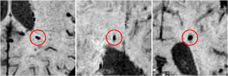
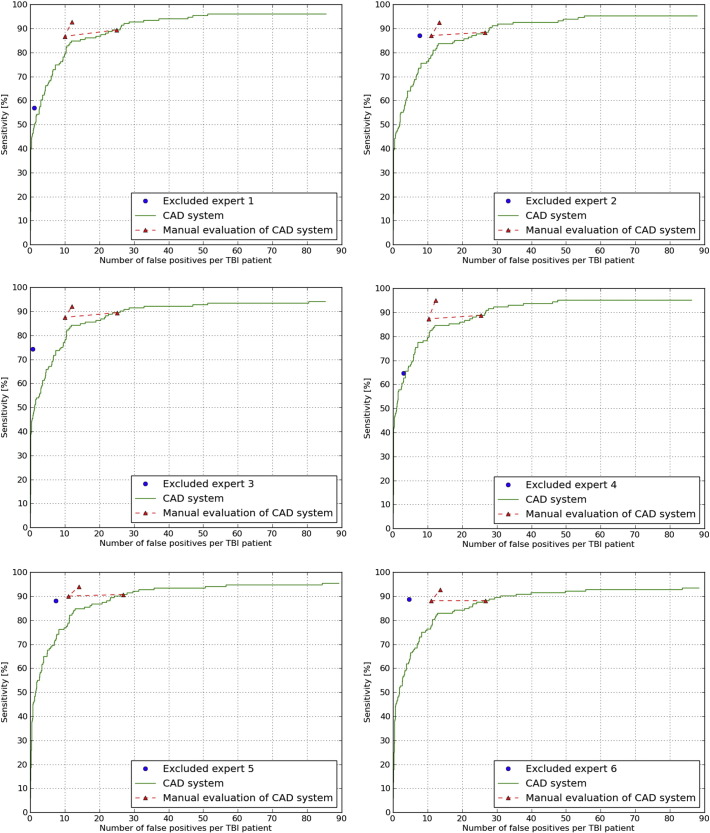
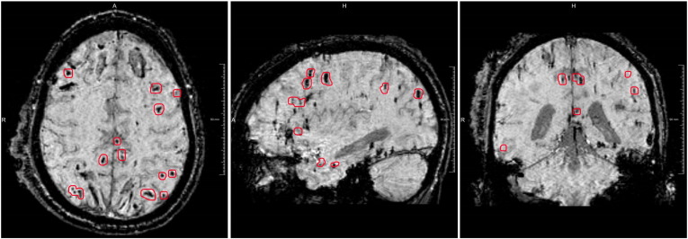
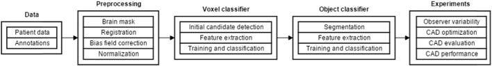
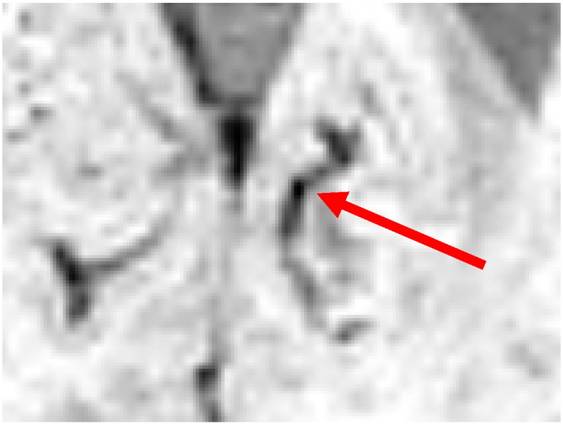
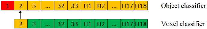
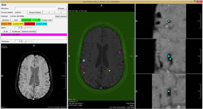
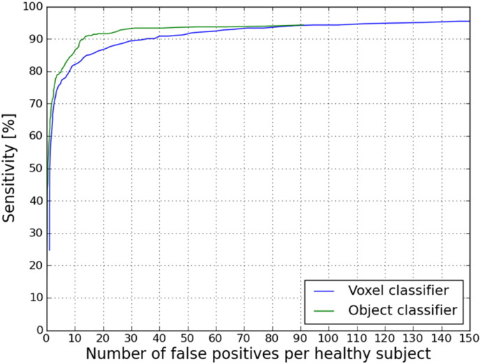
In this paper a Computer Aided Detection (CAD) system is presented to automatically detect Cerebral Microbleeds (CMBs) in patients with Traumatic Brain Injury (TBI). It is believed that the presence of CMBs has clinical prognostic value in TBI patients. To study the contribution of CMBs in patient outcome, accurate detection of CMBs is required. Manual detection of CMBs in TBI patients is a time consuming task that is prone to errors, because CMBs are easily overlooked and are difficult to distinguish from blood vessels. This study included 33 TBI patients. Because of the laborious nature of manually annotating CMBs, only one trained expert manually annotated the CMBs in all 33 patients. A subset of ten TBI patients was annotated by six experts. Our CAD system makes use of both Susceptibility Weighted Imaging (SWI) and T1 weighted magnetic resonance images to detect CMBs. After pre-processing these images, a two-step approach was used for automated detection of CMBs. In the first step, each voxel was characterized by twelve features based on the dark and spherical nature of CMBs and a random forest classifier was used to identify CMB candidate locations. In the second step, segmentations were made from each identified candidate location. Subsequently an object-based classifier was used to remove false positive detections of the voxel classifier, by considering seven object-based features that discriminate between spherical objects (CMBs) and elongated objects (blood vessels). A guided user interface was designed for fast evaluation of the CAD system result. During this process, an expert checked each CMB detected by the CAD system. A Fleiss' kappa value of only 0.24 showed that the inter-observer variability for the TBI patients in this study was very large. An expert using the guided user interface reached an average sensitivity of 93%, which was significantly higher (p = 0.03) than the average sensitivity of 77% (sd 12.4%) that the six experts manually detected. Furthermore, with the use of this CAD system the reading time was substantially reduced from one hour to 13 minutes per patient, because the CAD system only detects on average 25.9 false positives per TBI patient, resulting in 0.29 false positives per definite CMB finding.
本文提出了一种计算机辅助检测(CAD)系统,用于自动检测创伤性脑损伤(TBI)患者的脑微出血(CMB)。据信,CMB的存在对TBI患者具有临床预后价值。为了研究CMB对患者预后的影响,需要准确检测CMB。在TBI患者中手动检测CMB是一项耗时的任务,且容易出错,因为CMB很容易被忽视,并且难以与血管区分开来。本研究纳入了33例TBI患者。由于手动标注CMB的工作繁重,只有一位经过培训的专家对所有33例患者的CMB进行了手动标注。十例TBI患者的子集由六位专家进行了标注。我们的CAD系统利用了磁敏感加权成像(SWI)和T1加权磁共振图像来检测CMB。在对这些图像进行预处理后,采用两步法自动检测CMB。第一步,基于CMB的暗区和球形特征,用十二个特征对每个体素进行表征,并使用随机森林分类器识别CMB候选位置。第二步,从每个识别出的候选位置进行分割。随后,使用基于对象的分类器,通过考虑七个基于对象的特征来区分球形对象(CMB)和细长对象(血管),以去除体素分类器的假阳性检测。设计了一个引导式用户界面,用于快速评估CAD系统的结果。在此过程中,一位专家检查了CAD系统检测到的每个CMB。Fleiss卡方值仅为0.24,表明本研究中TBI患者的观察者间变异性非常大。使用引导式用户界面的一位专家达到了93%的平均敏感度,这显著高于六位专家手动检测时77%(标准差12.4%)的平均敏感度(p = 0.03)。此外,使用该CAD系统后,每位患者的阅片时间从一小时大幅减少至13分钟,因为CAD系统平均每位TBI患者仅检测到25.9例假阳性,即每确定一个CMB发现仅有0.29例假阳性。