Pan Peng, Zhang Pengsong, Premachandran Sharanja, Peng Ran, Wang Shaojia, Fan Qigao, Sun Yu, Calarco John A, Liu Xinyu
Department of Mechanical and Industrial Engineering, University of Toronto, Toronto, Ontario M5S 3G8, Canada.
Department of Cell & Systems Biology, University of Toronto, Toronto, Ontario M5S 3G5, Canada.
Research (Wash D C). 2024 Oct 30;7:0513. doi: 10.34133/research.0513. eCollection 2024.
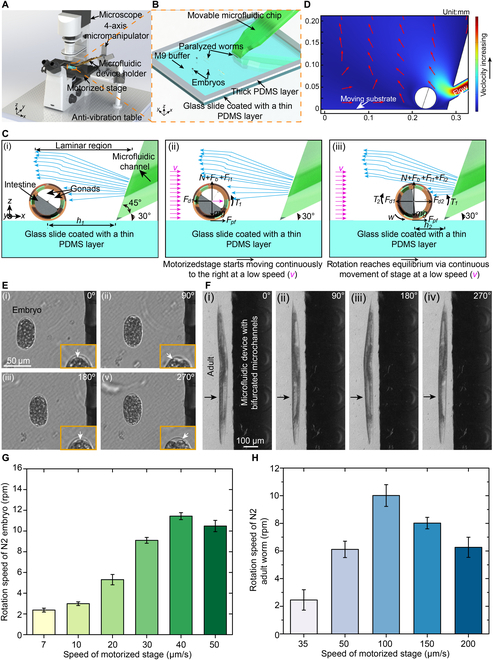
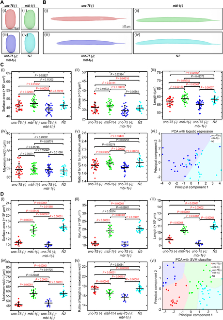
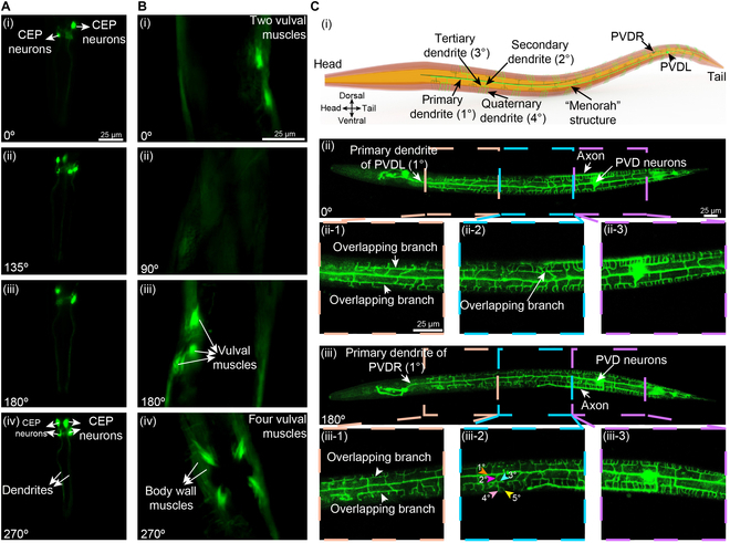
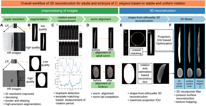
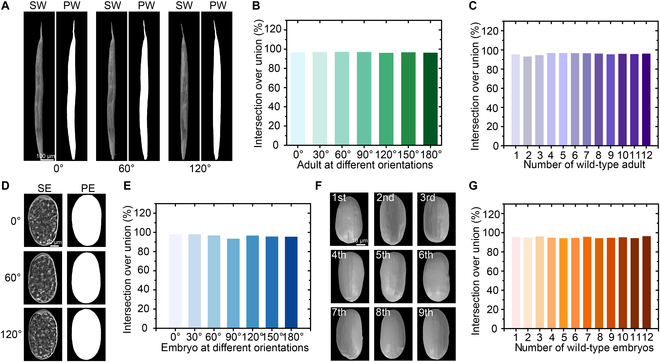
Accurate visualization and 3-dimensional (3D) morphological profiling of small model organisms can provide quantitative phenotypes benefiting genetic analysis and modeling of human diseases in tractable organisms. However, in the highly studied nematode accurate morphological phenotyping remains challenging because of notable decrease in image resolution of distant signal under high magnification and complexity in the 3D reconstruction of microscale samples with irregular shapes. Here, we develop a robust robotic system that enables the contactless, stable, and uniform rotation of for multi-view fluorescent imaging and 3D morphological phenotyping via the precise reconstruction of 3D models. Contactless animal rotation accommodates a variety of body shapes and sizes found at different developmental stages and in mutant strains. Through controlled rotation, high-resolution fluorescent imaging of structures is obtained by overcoming the limitations inherent in both widefield and confocal microscopy. Combining our robotic system with machine learning, we create, for the first time, precise 3D reconstructions of at the embryonic and adult stages, enabling 3D morphological phenotyping of mutant strains in an accurate and comprehensive fashion. Intriguingly, our morphological phenotyping discovered a genetic interaction between 2 RNA binding proteins (UNC-75/CELF and MBL-1/MBNL), which are highly conserved between and humans and implicated in neurological and muscular disorders. Our system can thus generate quantitative morphological readouts facilitating the investigation of genetic variations and disease mechanisms. More broadly, our method will also be amenable for 3D phenotypic analysis of other biological samples, like zebrafish and larvae.
对小型模式生物进行精确可视化和三维(3D)形态学分析,可为遗传学分析以及在易处理生物中对人类疾病进行建模提供定量表型。然而,在被广泛研究的线虫中,由于高倍放大下远距离信号的图像分辨率显著降低,以及对形状不规则的微观样本进行三维重建时存在复杂性,精确的形态学表型分析仍然具有挑战性。在此,我们开发了一种强大的机器人系统,该系统能够通过精确重建三维模型,实现线虫的非接触式、稳定且均匀的旋转,以进行多视角荧光成像和三维形态学表型分析。非接触式动物旋转适用于不同发育阶段和突变株中发现的各种体型和大小。通过可控旋转,克服了广角显微镜和共聚焦显微镜固有的局限性,获得了线虫结构的高分辨率荧光成像。将我们的机器人系统与机器学习相结合,我们首次创建了线虫胚胎期和成虫期的精确三维重建,能够以准确和全面的方式对线虫突变株进行三维形态学表型分析。有趣的是,我们的形态学表型分析发现了两种RNA结合蛋白(UNC - 75/CELF和MBL - 1/MBNL)之间的遗传相互作用,这两种蛋白在线虫和人类之间高度保守,并与神经和肌肉疾病有关。因此,我们的系统可以生成定量的形态学读数,便于研究遗传变异和疾病机制。更广泛地说,我们的方法也适用于对其他生物样本(如斑马鱼和果蝇幼虫)进行三维表型分析。