Smetanick Derek, Stolley Danielle, Fuentes David, Fowlkes Natalie W, Shakoor Faith, Stenkamp Maria Sophia, Hicks Samantha, Parrish Steve, Cressman Erik
University of Arizona College of Medicine Tucson, Tucson, AZ 85724, USA.
Department of Interventional Radiology, MD Anderson Cancer Center, The University of Texas, 1515 Holcombe Blvd., Houston, TX 77030, USA.
Life (Basel). 2024 Oct 30;14(11):1395. doi: 10.3390/life14111395.
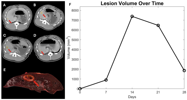
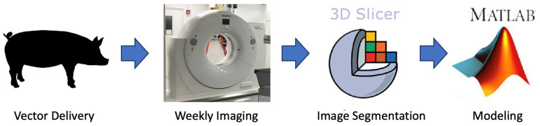
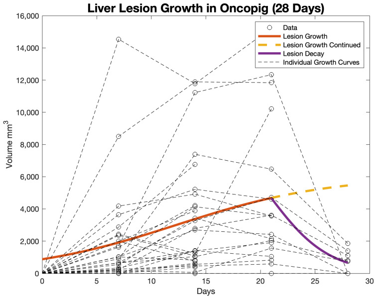
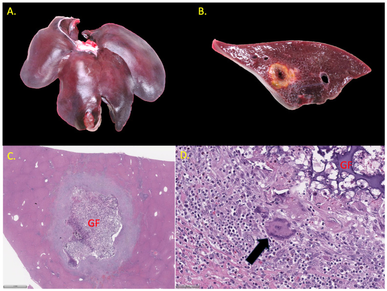
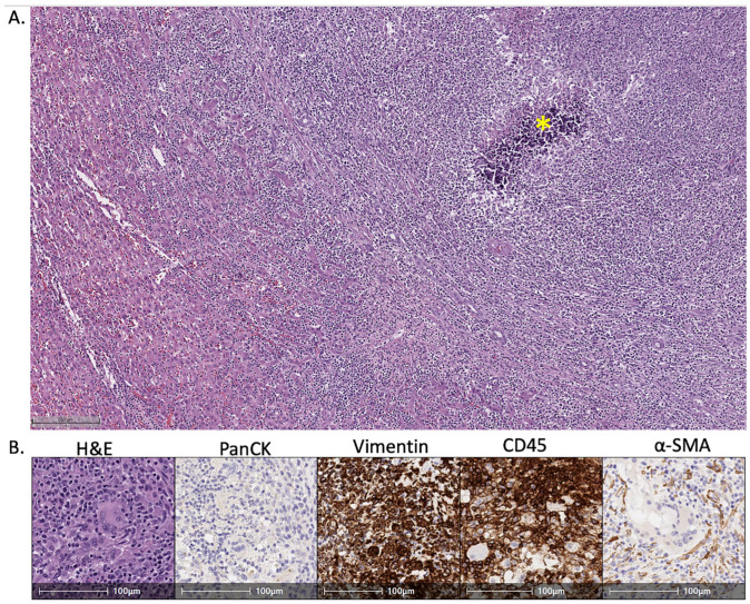
The growth rate of in situ-induced hepatic lesions in an Oncopig large animal model is quantitatively assessed. Oncopigs (n = 9) received baseline triple-phase CT scans prior to lesion induction. Lesions were subsequently induced by delivering the Ad-Cre vector to four locations in the liver. Triple-phase CT scans were obtained weekly to track the growth of the lesions. Animals were sacrificed at 14, 21, or 28 days (n = 3 in each group). The overall success rate of lesion generation was ~78%. Histopathology sections consistently revealed lesions that were highly inflammatory and consisted of a large leukocyte population without clear evidence of carcinomas. Lesions presented within imaging as hypovascular, low attenuating masses with slight contrast enhancement around the margins but little to no enhancement within the lesions themselves. The observed lesions were manually segmented on the venous phase image. Segmentation volumes were fitted to a logistic growth and decay model. Several lesions observed at earlier time points in the 28-day group had fully regressed by the time of the necropsy. The overall trend of rapid growth for the first 21 days, with spontaneous regression of the lesions being observed from day 21 to 28, suggests that the optimal window for experimental studies may be from days 14 to 21. The data and mathematical models generated from this study may be used for future computational models; however, the current model presented has moderate clinical relevance because many induced tumors resolved spontaneously within a few weeks. Awareness and careful consideration of the modest relevance and limitations of the model are advisable for each specific use case.
在一种肿瘤猪大型动物模型中,对原位诱导的肝脏病变的生长速率进行了定量评估。肿瘤猪(n = 9)在病变诱导前接受了基线三期CT扫描。随后通过将Ad-Cre载体递送至肝脏的四个位置来诱导病变。每周进行三期CT扫描以追踪病变的生长情况。在14天、21天或28天时对动物实施安乐死(每组n = 3)。病变生成的总体成功率约为78%。组织病理学切片始终显示病变具有高度炎症性,由大量白细胞组成,无明显的癌证据。病变在影像学上表现为低血供、低衰减肿块,边缘有轻微的对比增强,但病变内部几乎没有增强。在静脉期图像上对观察到的病变进行手动分割。分割体积拟合到逻辑生长和衰减模型。在28天组中,在较早时间点观察到的几个病变在尸检时已完全消退。前21天快速生长的总体趋势,以及从第21天到第28天观察到病变自发消退,表明实验研究的最佳窗口可能是从第14天到第21天。本研究生成的数据和数学模型可用于未来的计算模型;然而,目前提出的模型具有中等临床相关性,因为许多诱导肿瘤在几周内自发消退。对于每个具体的使用案例,建议了解并仔细考虑该模型适度的相关性和局限性。