• 文献检索
  • 文档翻译
  • 深度研究
  • 学术资讯
  • Suppr Zotero 插件Zotero 插件
  • 邀请有礼
  • 套餐&价格
  • 历史记录
应用&插件
Suppr Zotero 插件Zotero 插件浏览器插件Mac 客户端Windows 客户端微信小程序
定价
高级版会员购买积分包购买API积分包
服务
文献检索文档翻译深度研究API 文档MCP 服务
关于我们
关于 Suppr公司介绍联系我们用户协议隐私条款
关注我们

Suppr 超能文献

核心技术专利:CN118964589B侵权必究
粤ICP备2023148730 号-1Suppr @ 2026

文献检索

告别复杂PubMed语法,用中文像聊天一样搜索,搜遍4000万医学文献。AI智能推荐,让科研检索更轻松。

立即免费搜索

文件翻译

保留排版,准确专业,支持PDF/Word/PPT等文件格式,支持 12+语言互译。

免费翻译文档

深度研究

AI帮你快速写综述,25分钟生成高质量综述,智能提取关键信息,辅助科研写作。

立即免费体验

利用深度迁移学习在异质性计算机断层扫描图像中自动检测和分割颈部淋巴结

Automatic cervical lymph nodes detection and segmentation in heterogeneous computed tomography images using deep transfer learning.

作者信息

Liao Wenjun, Luo Xiangde, Li Lu, Xu Jinfeng, He Yuan, Huang Hui, Zhang Shichuan

机构信息

Department of Radiation Oncology, Sichuan Cancer Hospital and Institute, Sichuan Cancer Center, Cancer Hospital Affiliate to School of Medicine, University of Electronic Science and Technology of China, Chengdu, 610041, China.

School of Mechanical and Electrical Engineering, University of Electronic Science and Technology of China, Chengdu, 611731, China.

出版信息

Sci Rep. 2025 Feb 4;15(1):4250. doi: 10.1038/s41598-024-84804-3.

DOI:10.1038/s41598-024-84804-3
PMID:39905029
原文链接:https://pmc.ncbi.nlm.nih.gov/articles/PMC11794882/
Abstract

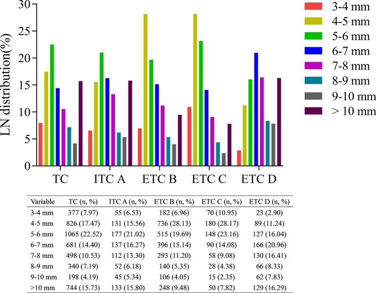
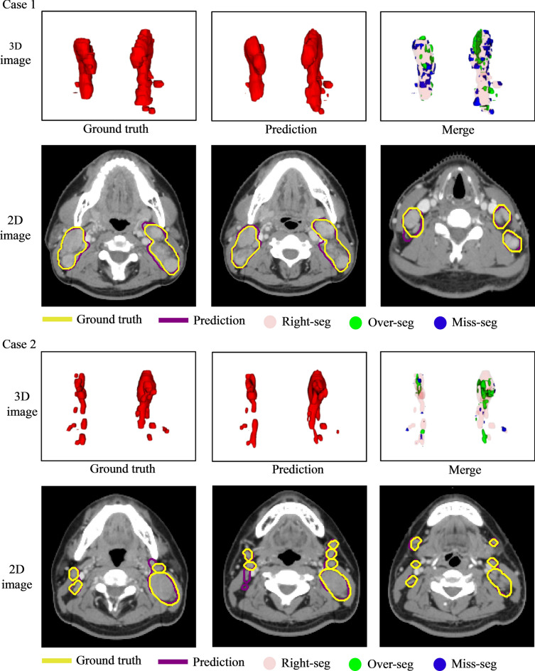
To develop a deep learning model using transfer learning for automatic detection and segmentation of neck lymph nodes (LNs) in computed tomography (CT) images, the study included 11,013 annotated LNs with a short-axis diameter ≥ 3 mm from 626 head and neck cancer patients across four hospitals. The nnUNet model was used as a baseline, pre-trained on a large-scale head and neck dataset, and then fine-tuned with 4,729 LNs from hospital A for detection and segmentation. Validation was conducted on an internal testing cohort (ITC A) and three external testing cohorts (ETCs B, C, and D), with 1684 and 4600 LNs, respectively. Detection was evaluated via sensitivity, positive predictive value (PPV), and false positive rate per case (FP/vol), while segmentation was assessed using the Dice similarity coefficient (DSC) and Hausdorff distance (HD95). For detection, the sensitivity, PPV, and FP/vol in ITC A were 54.6%, 69.0%, and 3.4, respectively. In ETCs, the sensitivity ranged from 45.7% at 3.9 FP/vol to 63.5% at 5.8 FP/vol. Segmentation achieved a mean DSC of 0.72 in ITC A and 0.72 to 0.74 in ETCs, as well as a mean HD95 of 3.78 mm in ITC A and 2.73 mm to 2.85 mm in ETCs. No significant sensitivity difference was found between contrast-enhanced and unenhanced CT images (p = 0.502) or repeated CT images (p = 0.815) during adaptive radiotherapy. The model's segmentation accuracy was comparable to that of experienced oncologists. The model shows promise in automatically detecting and segmenting neck LNs in CT images, potentially reducing oncologists' segmentation workload.

摘要

为了开发一种使用迁移学习的深度学习模型,用于在计算机断层扫描(CT)图像中自动检测和分割颈部淋巴结(LN),该研究纳入了来自四家医院的626例头颈癌患者的11,013个短轴直径≥3mm的标注淋巴结。nnUNet模型被用作基线,在大规模头颈数据集上进行预训练,然后使用医院A的4,729个淋巴结进行微调以进行检测和分割。在内部测试队列(ITC A)和三个外部测试队列(ETC B、C和D)上进行验证,分别有1684个和4600个淋巴结。通过灵敏度、阳性预测值(PPV)和每例假阳性率(FP/vol)评估检测,而使用Dice相似系数(DSC)和豪斯多夫距离(HD95)评估分割。对于检测,ITC A中的灵敏度、PPV和FP/vol分别为54.6%、69.0%和3.4。在ETC中,灵敏度范围从FP/vol为3.9时的45.7%到FP/vol为5.8时的63.5%。分割在ITC A中平均DSC为0.72,在ETC中为0.72至0.74,ITC A中的平均HD95为3.78mm,ETC中为2.73mm至2.85mm。在自适应放疗期间,对比增强CT图像和未增强CT图像之间(p = 0.502)或重复CT图像之间(p = 0.815)未发现显著的灵敏度差异。该模型的分割准确性与经验丰富的肿瘤学家相当。该模型在自动检测和分割CT图像中的颈部淋巴结方面显示出前景,可能会减少肿瘤学家的分割工作量。

https://cdn.ncbi.nlm.nih.gov/pmc/blobs/e91f/11794882/24e012782ac7/41598_2024_84804_Fig6_HTML.jpg
https://cdn.ncbi.nlm.nih.gov/pmc/blobs/e91f/11794882/839e65b5ac6c/41598_2024_84804_Fig1_HTML.jpg
https://cdn.ncbi.nlm.nih.gov/pmc/blobs/e91f/11794882/188c46d3ca87/41598_2024_84804_Fig2_HTML.jpg
https://cdn.ncbi.nlm.nih.gov/pmc/blobs/e91f/11794882/7cee0f184ad7/41598_2024_84804_Fig3_HTML.jpg
https://cdn.ncbi.nlm.nih.gov/pmc/blobs/e91f/11794882/d8c5ca986a0b/41598_2024_84804_Fig4_HTML.jpg
https://cdn.ncbi.nlm.nih.gov/pmc/blobs/e91f/11794882/9142ee663c1a/41598_2024_84804_Fig5_HTML.jpg
https://cdn.ncbi.nlm.nih.gov/pmc/blobs/e91f/11794882/24e012782ac7/41598_2024_84804_Fig6_HTML.jpg

相似文献

1
Automatic cervical lymph nodes detection and segmentation in heterogeneous computed tomography images using deep transfer learning.利用深度迁移学习在异质性计算机断层扫描图像中自动检测和分割颈部淋巴结
Sci Rep. 2025 Feb 4;15(1):4250. doi: 10.1038/s41598-024-84804-3.
2
Deep learning-based fully automated detection and segmentation of pelvic lymph nodes on diffusion-weighted images for prostate cancer: a multicenter study.基于深度学习的前列腺癌扩散加权图像上盆腔淋巴结的全自动检测与分割:一项多中心研究
Cancer Imaging. 2025 Mar 17;25(1):37. doi: 10.1186/s40644-025-00840-w.
3
Deep learning-based fully automated detection and segmentation of lymph nodes on multiparametric-mri for rectal cancer: A multicentre study.基于深度学习的直肠癌多参数 MRI 淋巴结全自动检测与分割:一项多中心研究。
EBioMedicine. 2020 Jun;56:102780. doi: 10.1016/j.ebiom.2020.102780. Epub 2020 Jun 5.
4
Multi-modal segmentation with missing image data for automatic delineation of gross tumor volumes in head and neck cancers.多模态分割中存在图像缺失数据的情况下,实现头颈部癌症大体肿瘤体积的自动勾画。
Med Phys. 2024 Oct;51(10):7295-7307. doi: 10.1002/mp.17260. Epub 2024 Jun 19.
5
Automated detection and segmentation of thoracic lymph nodes from CT using 3D foveal fully convolutional neural networks.基于三维焦域全卷积神经网络的 CT 图像胸部淋巴结自动检测与分割
BMC Med Imaging. 2021 Apr 13;21(1):69. doi: 10.1186/s12880-021-00599-z.
6
Comparison of deep learning networks for fully automated head and neck tumor delineation on multi-centric PET/CT images.多中心 PET/CT 图像上全自动头颈部肿瘤勾画的深度学习网络比较。
Radiat Oncol. 2024 Jan 8;19(1):3. doi: 10.1186/s13014-023-02388-0.
7
Automated localization and segmentation of cervical lymph nodes on contrast-enhanced CT using a 3D foveal fully convolutional neural network.使用三维焦点全卷积神经网络对增强 CT 上的颈部淋巴结进行自动定位和分割。
Eur Radiol Exp. 2023 Jul 28;7(1):45. doi: 10.1186/s41747-023-00360-x.
8
Automated Segmentation of Lymph Nodes on Neck CT Scans Using Deep Learning.利用深度学习对颈部CT扫描图像中的淋巴结进行自动分割
J Imaging Inform Med. 2024 Dec;37(6):2955-2966. doi: 10.1007/s10278-024-01114-w. Epub 2024 Jun 27.
9
Deep learning vs. atlas-based models for fast auto-segmentation of the masticatory muscles on head and neck CT images.深度学习与基于图谱的模型在头颈部 CT 图像咀嚼肌自动分割中的比较。
Radiat Oncol. 2020 Jul 20;15(1):176. doi: 10.1186/s13014-020-01617-0.
10
Application of deep-learning to the automatic segmentation and classification of lateral lymph nodes on ultrasound images of papillary thyroid carcinoma.深度学习在甲状腺乳头状癌超声图像中侧颈淋巴结自动分割和分类的应用。
Asian J Surg. 2024 Sep;47(9):3892-3898. doi: 10.1016/j.asjsur.2024.02.140. Epub 2024 Mar 6.

引用本文的文献

1
Deep learning for malignant lymph node segmentation and detection: a review.深度学习在恶性淋巴结分割与检测中的应用综述
Front Immunol. 2025 Apr 28;16:1526518. doi: 10.3389/fimmu.2025.1526518. eCollection 2025.

本文引用的文献

1
SegRap2023: A benchmark of organs-at-risk and gross tumor volume Segmentation for Radiotherapy Planning of Nasopharyngeal Carcinoma.SegRap2023:鼻咽癌放疗计划中危及器官和大体肿瘤体积分割的基准
Med Image Anal. 2025 Apr;101:103447. doi: 10.1016/j.media.2024.103447. Epub 2025 Jan 2.
2
LLM-driven multimodal target volume contouring in radiation oncology.人工智能驱动的多模态靶区勾画在放射肿瘤学中的应用。
Nat Commun. 2024 Oct 24;15(1):9186. doi: 10.1038/s41467-024-53387-y.
3
Use of Pretreatment Perfusion MRI-based Intratumoral Heterogeneity to Predict Pathologic Response of Triple-Negative Breast Cancer to Neoadjuvant Chemoimmunotherapy.
使用预处理灌注 MRI 评估肿瘤内异质性预测三阴性乳腺癌新辅助化疗免疫治疗的病理反应。
Radiology. 2024 Sep;312(3):e240575. doi: 10.1148/radiol.240575.
4
Multiregional dynamic contrast-enhanced MRI-based integrated system for predicting pathological complete response of axillary lymph node to neoadjuvant chemotherapy in breast cancer: multicentre study.多区域动态对比增强 MRI 为基础的预测乳腺癌腋窝淋巴结新辅助化疗病理完全缓解的集成系统:多中心研究。
EBioMedicine. 2024 Sep;107:105311. doi: 10.1016/j.ebiom.2024.105311. Epub 2024 Aug 26.
5
A multicenter clinical AI system study for detection and diagnosis of focal liver lesions.多中心临床人工智能系统研究用于检测和诊断局灶性肝病变。
Nat Commun. 2024 Feb 7;15(1):1131. doi: 10.1038/s41467-024-45325-9.
6
Evaluation of mediastinal lymph node segmentation of heterogeneous CT data with full and weak supervision.使用全监督和弱监督方法评估异质 CT 数据的纵隔淋巴结分割。
Comput Med Imaging Graph. 2024 Jan;111:102312. doi: 10.1016/j.compmedimag.2023.102312. Epub 2023 Dec 15.
7
LNAS: a clinically applicable deep-learning system for mediastinal enlarged lymph nodes segmentation and station mapping without regard to the pathogenesis using unenhanced CT images.LNAS:一种临床适用的深度学习系统,用于在不考虑发病机制的情况下,使用未增强 CT 图像对纵隔增大淋巴结进行分割和定位。
Radiol Med. 2024 Feb;129(2):229-238. doi: 10.1007/s11547-023-01747-x. Epub 2023 Dec 18.
8
Development and Evaluation of Deep Learning Models for Automated Estimation of Myelin Maturation Using Pediatric Brain MRI Scans.利用儿科脑磁共振成像扫描进行髓鞘成熟度自动估计的深度学习模型的开发与评估
Radiol Artif Intell. 2023 Jul 26;5(5):e220292. doi: 10.1148/ryai.220292. eCollection 2023 Sep.
9
TotalSegmentator: Robust Segmentation of 104 Anatomic Structures in CT Images.全段分割器:CT图像中104种解剖结构的稳健分割
Radiol Artif Intell. 2023 Jul 5;5(5):e230024. doi: 10.1148/ryai.230024. eCollection 2023 Sep.
10
Automated localization and segmentation of cervical lymph nodes on contrast-enhanced CT using a 3D foveal fully convolutional neural network.使用三维焦点全卷积神经网络对增强 CT 上的颈部淋巴结进行自动定位和分割。
Eur Radiol Exp. 2023 Jul 28;7(1):45. doi: 10.1186/s41747-023-00360-x.