• 文献检索
  • 文档翻译
  • 深度研究
  • 学术资讯
  • Suppr Zotero 插件Zotero 插件
  • 邀请有礼
  • 套餐&价格
  • 历史记录
应用&插件
Suppr Zotero 插件Zotero 插件浏览器插件Mac 客户端Windows 客户端微信小程序
定价
高级版会员购买积分包购买API积分包
服务
文献检索文档翻译深度研究API 文档MCP 服务
关于我们
关于 Suppr公司介绍联系我们用户协议隐私条款
关注我们

Suppr 超能文献

核心技术专利:CN118964589B侵权必究
粤ICP备2023148730 号-1Suppr @ 2026

文献检索

告别复杂PubMed语法,用中文像聊天一样搜索,搜遍4000万医学文献。AI智能推荐,让科研检索更轻松。

立即免费搜索

文件翻译

保留排版,准确专业,支持PDF/Word/PPT等文件格式,支持 12+语言互译。

免费翻译文档

深度研究

AI帮你快速写综述,25分钟生成高质量综述,智能提取关键信息,辅助科研写作。

立即免费体验

使用扫描激光检眼镜观察眼底自发荧光的分布情况。

Distribution of fundus autofluorescence with a scanning laser ophthalmoscope.

作者信息

von Rückmann A, Fitzke F W, Bird A C

机构信息

Institute of Ophthalmology, Moorfields Eye Hospital, London.

出版信息

Br J Ophthalmol. 1995 May;79(5):407-12. doi: 10.1136/bjo.79.5.407.

DOI:10.1136/bjo.79.5.407
PMID:7612549
原文链接:https://pmc.ncbi.nlm.nih.gov/articles/PMC505125/
Abstract

BACKGROUND

Variation of fluorescence derived from lipofuscin in the retinal pigment epithelium has been recorded with age and in retinal diseases. Studies have been based largely on in vitro observations on eye bank eyes which has placed severe limitations on the data available.

METHODS

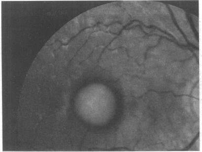
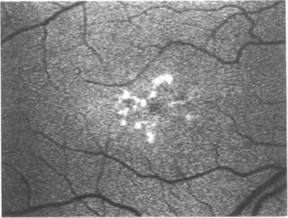
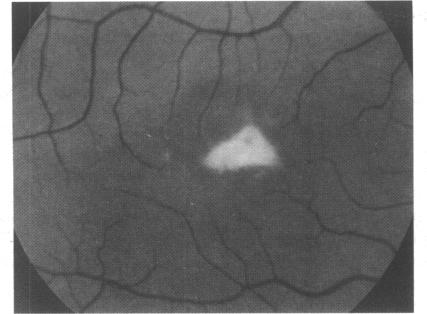
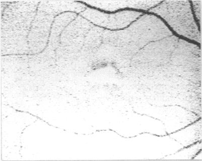
A technique is described whereby in vivo imaging of autofluorescence of the fundus was achieved using a scanning laser ophthalmoscope.

RESULTS

The optical characteristics, distribution, and variation with disease imply that the fluorescence is derived from lipofuscin in the pigment epithelium. Autofluorescence is shown to be abnormally high in certain inherited diseases, and low in the presence of retinal atrophy.

CONCLUSION

This technique may be useful both in clinical practice and research. It may allow the detection of the abnormal phenotype in genetically determined disease at a time when other techniques may not. Longitudinal studies of age related macular disease would permit correlation between changes in the pigment epithelium and Bruch's membrane to be established.

摘要

背景

视网膜色素上皮中脂褐素产生的荧光变化已随年龄增长及在视网膜疾病中被记录下来。此前的研究很大程度上基于对眼库眼球的体外观察,这对可得数据造成了严重限制。

方法

本文描述了一种技术,通过该技术利用扫描激光检眼镜实现了眼底自发荧光的体内成像。

结果

其光学特性、分布以及随疾病的变化表明,这种荧光源自色素上皮中的脂褐素。自发荧光在某些遗传性疾病中显示异常高,而在存在视网膜萎缩时则较低。

结论

该技术在临床实践和研究中可能都有用。它可能在其他技术无法做到的时候检测出基因决定疾病中的异常表型。对年龄相关性黄斑疾病的纵向研究将有助于建立色素上皮和布鲁赫膜变化之间的关联。

https://cdn.ncbi.nlm.nih.gov/pmc/blobs/4afb/505125/8d9098b73c68/brjopthal00017-0013-c.jpg
https://cdn.ncbi.nlm.nih.gov/pmc/blobs/4afb/505125/ef9a4e608c4d/brjopthal00017-0009-a.jpg
https://cdn.ncbi.nlm.nih.gov/pmc/blobs/4afb/505125/f8bf717ca519/brjopthal00017-0010-a.jpg
https://cdn.ncbi.nlm.nih.gov/pmc/blobs/4afb/505125/f91fb73fa06d/brjopthal00017-0010-b.jpg
https://cdn.ncbi.nlm.nih.gov/pmc/blobs/4afb/505125/ea00d7137280/brjopthal00017-0010-c.jpg
https://cdn.ncbi.nlm.nih.gov/pmc/blobs/4afb/505125/a04709a700a1/brjopthal00017-0010-d.jpg
https://cdn.ncbi.nlm.nih.gov/pmc/blobs/4afb/505125/c48e6424bde9/brjopthal00017-0011-a.jpg
https://cdn.ncbi.nlm.nih.gov/pmc/blobs/4afb/505125/d71c3df31f80/brjopthal00017-0011-b.jpg
https://cdn.ncbi.nlm.nih.gov/pmc/blobs/4afb/505125/f7aad959e71c/brjopthal00017-0011-c.jpg
https://cdn.ncbi.nlm.nih.gov/pmc/blobs/4afb/505125/1592a9a49264/brjopthal00017-0011-d.jpg
https://cdn.ncbi.nlm.nih.gov/pmc/blobs/4afb/505125/3e86d139f82d/brjopthal00017-0012-a.jpg
https://cdn.ncbi.nlm.nih.gov/pmc/blobs/4afb/505125/27fe70279774/brjopthal00017-0012-b.jpg
https://cdn.ncbi.nlm.nih.gov/pmc/blobs/4afb/505125/2c1f28b61ce2/brjopthal00017-0012-c.jpg
https://cdn.ncbi.nlm.nih.gov/pmc/blobs/4afb/505125/f217a8a443c4/brjopthal00017-0012-d.jpg
https://cdn.ncbi.nlm.nih.gov/pmc/blobs/4afb/505125/424854c1b205/brjopthal00017-0012-e.jpg
https://cdn.ncbi.nlm.nih.gov/pmc/blobs/4afb/505125/4d471461d7f1/brjopthal00017-0013-a.jpg
https://cdn.ncbi.nlm.nih.gov/pmc/blobs/4afb/505125/d12f75475628/brjopthal00017-0013-b.jpg
https://cdn.ncbi.nlm.nih.gov/pmc/blobs/4afb/505125/8d9098b73c68/brjopthal00017-0013-c.jpg

相似文献

1
Distribution of fundus autofluorescence with a scanning laser ophthalmoscope.使用扫描激光检眼镜观察眼底自发荧光的分布情况。
Br J Ophthalmol. 1995 May;79(5):407-12. doi: 10.1136/bjo.79.5.407.
2
Fundus autofluorescence in age-related macular disease imaged with a laser scanning ophthalmoscope.使用激光扫描检眼镜对年龄相关性黄斑疾病进行眼底自发荧光成像。
Invest Ophthalmol Vis Sci. 1997 Feb;38(2):478-86.
3
Distribution of pigment epithelium autofluorescence in retinal disease state recorded in vivo and its change over time.视网膜疾病状态下色素上皮细胞自发荧光的活体记录分布及其随时间的变化。
Graefes Arch Clin Exp Ophthalmol. 1999 Jan;237(1):1-9. doi: 10.1007/s004170050186.
4
Patterns of increased in vivo fundus autofluorescence in the junctional zone of geographic atrophy of the retinal pigment epithelium associated with age-related macular degeneration.与年龄相关性黄斑变性相关的视网膜色素上皮地图状萎缩交界区体内眼底自发荧光增加的模式
Graefes Arch Clin Exp Ophthalmol. 1999 Feb;237(2):145-52. doi: 10.1007/s004170050209.
5
[Topography of fundus autofluorescence with a new confocal scanning laser ophthalmoscope].
Ophthalmologe. 1997 Jun;94(6):385-91. doi: 10.1007/s003470050130.
6
In vivo fundus autofluorescence in macular dystrophies.黄斑营养不良的活体眼底自发荧光
Arch Ophthalmol. 1997 May;115(5):609-15. doi: 10.1001/archopht.1997.01100150611006.
7
Quantitative evaluation of fundus autofluorescence imaged "in vivo" in eyes with retinal disease.视网膜疾病患者眼部“活体”眼底自发荧光成像的定量评估。
Br J Ophthalmol. 2000 Jul;84(7):741-5. doi: 10.1136/bjo.84.7.741.
8
Lack of fundus autofluorescence to 488 nanometers from childhood on in patients with early-onset severe retinal dystrophy associated with mutations in RPE65.患有与RPE65基因突变相关的早发性严重视网膜营养不良的患者自儿童期起缺乏对488纳米的眼底自发荧光。
Ophthalmology. 2004 Aug;111(8):1585-94. doi: 10.1016/j.ophtha.2004.01.033.
9
Fundus autofluorescence and development of geographic atrophy in age-related macular degeneration.年龄相关性黄斑变性中的眼底自发荧光与地图样萎缩的发展
Invest Ophthalmol Vis Sci. 2001 Apr;42(5):1051-6.
10
Fundus autofluorescence imaging compared with different confocal scanning laser ophthalmoscopes.使用不同的共焦扫描激光眼科显微镜进行眼底自发荧光成像比较。
Br J Ophthalmol. 2003 Nov;87(11):1381-6. doi: 10.1136/bjo.87.11.1381.

引用本文的文献

1
Stargardt's Disease: Molecular Pathogenesis and Current Therapeutic Landscape.斯塔加特病:分子发病机制与当前治疗现状
Int J Mol Sci. 2025 Jul 21;26(14):7006. doi: 10.3390/ijms26147006.
2
Fundus Autofluorescence in Inherited Retinal Disease: A Review.遗传性视网膜疾病中的眼底自发荧光:综述
Cells. 2025 Jul 16;14(14):1092. doi: 10.3390/cells14141092.
3
Blue Light and Green Light Fundus Autofluorescence, Complementary to Optical Coherence Tomography, in Age-Related Macular Degeneration Evaluation.蓝光和绿光眼底自发荧光在年龄相关性黄斑变性评估中对光学相干断层扫描的补充作用

本文引用的文献

1
Heredodegeneration of the macula lutea; a study of the clinical and pathologic aspects.黄斑部遗传性变性;临床与病理方面的研究
AMA Arch Ophthalmol. 1955 Feb;53(2):224-8. doi: 10.1001/archopht.1955.00930010226009.
2
Mutations in the human retinal degeneration slow (RDS) gene can cause either retinitis pigmentosa or macular dystrophy.人类视网膜变性慢(RDS)基因的突变可导致色素性视网膜炎或黄斑营养不良。
Nat Genet. 1993 Mar;3(3):213-8. doi: 10.1038/ng0393-213.
3
Correlation between lipids extracted from Bruch's membrane and age.
Diagnostics (Basel). 2025 Jul 2;15(13):1688. doi: 10.3390/diagnostics15131688.
4
Bisretinoid lipofuscin, fundus autofluorescence and retinal disease.双视黄醛脂褐质、眼底自发荧光与视网膜疾病。
Prog Retin Eye Res. 2025 Jul 8;108:101388. doi: 10.1016/j.preteyeres.2025.101388.
5
Discrepancies Between Autofluorescence Imaging Modalities in CNGB3-Associated Achromatopsia and Correlation With Ellipsoid Zone Continuity.CNGB3相关性全色盲中自体荧光成像模式的差异及其与椭圆体区连续性的相关性
Transl Vis Sci Technol. 2025 May 1;14(5):24. doi: 10.1167/tvst.14.5.24.
6
Geographic atrophy: Understanding the relationship between structure and function.地图样萎缩:理解结构与功能之间的关系。
Asia Pac J Ophthalmol (Phila). 2025 May-Jun;14(3):100207. doi: 10.1016/j.apjo.2025.100207. Epub 2025 May 19.
7
Extent of Complete Retinal Pigment Epithelial and Outer Retinal Atrophy with Foveal Center Involvement is Associated with Visual Acuity.伴有黄斑中心凹受累的完全性视网膜色素上皮和外层视网膜萎缩程度与视力相关。
Ophthalmol Sci. 2024 Aug 29;5(1):100612. doi: 10.1016/j.xops.2024.100612. eCollection 2025 Jan-Feb.
8
Maculopathies: A Systematic Literature Review on Pathophysiology, Public Health, and Treatment.黄斑病变:关于病理生理学、公共卫生与治疗的系统文献综述
Cureus. 2024 Dec 1;16(12):e74911. doi: 10.7759/cureus.74911. eCollection 2024 Dec.
9
Identifying retinal pigment epithelium cells in adaptive optics-optical coherence tomography images with partial annotations and superhuman accuracy.在具有部分标注和超人精度的自适应光学光学相干断层扫描图像中识别视网膜色素上皮细胞。
Biomed Opt Express. 2024 Nov 21;15(12):6922-6939. doi: 10.1364/BOE.538473. eCollection 2024 Dec 1.
10
OCT analysis and MPOD assessment in patients affected by retinitis pigmentosa.OCT 分析和 MPOD 评估在视网膜色素变性患者中的应用。
Sci Rep. 2024 Nov 21;14(1):28830. doi: 10.1038/s41598-024-79979-8.
Ophthalmology. 1993 Jan;100(1):47-51. doi: 10.1016/s0161-6420(13)31712-6.
4
Three-dimensional visualization of confocal sections of in vivo human fundus and optic nerve.
Curr Eye Res. 1993 Nov;12(11):1015-8. doi: 10.3109/02713689309029228.
5
Analysis of lipid deposits extracted from human macular and peripheral Bruch's membrane.对从人黄斑和周边布鲁赫膜中提取的脂质沉积物的分析。
Arch Ophthalmol. 1994 Mar;112(3):402-6. doi: 10.1001/archopht.1994.01090150132035.
6
Histopathology and immunocytochemistry of the neurosensory retina in fundus flavimaculatus.
Ophthalmology. 1994 Jul;101(7):1211-9. doi: 10.1016/s0161-6420(13)31725-4.
7
A histopathologic study of Best's macular dystrophy.贝斯特黄斑营养不良的组织病理学研究。
Arch Ophthalmol. 1982 Jul;100(7):1115-21. doi: 10.1001/archopht.1982.01030040093017.
8
Histopathology of Best's macular dystrophy.贝斯特黄斑营养不良的组织病理学
Arch Ophthalmol. 1982 Jul;100(7):1108-14. doi: 10.1001/archopht.1982.01030040086016.
9
Two forms of autosomal dominant primary retinitis pigmentosa.常染色体显性遗传性原发性视网膜色素变性的两种类型。
Doc Ophthalmol. 1981 Nov;51(4):289-346. doi: 10.1007/BF00143336.
10
Rod and cone activity in patients with dominantly inherited retinitis pigmentosa: comparisons between psychophysical and electroretinographic measurements.常染色体显性遗传性视网膜色素变性患者的视杆和视锥细胞活性:心理物理学与视网膜电图测量结果的比较
Br J Ophthalmol. 1983 Jul;67(7):405-18. doi: 10.1136/bjo.67.7.405.