Seko Y, Minota S, Kawasaki A, Shinkai Y, Maeda K, Yagita H, Okumura K, Sato O, Takagi A, Tada Y
Third Department of Internal Medicine, Faculty of Medicine, University of Tokyo, Japan.
J Clin Invest. 1994 Feb;93(2):750-8. doi: 10.1172/JCI117029.
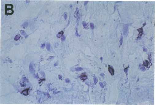
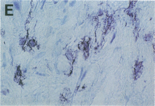
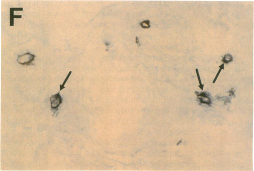
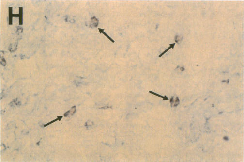
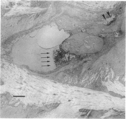
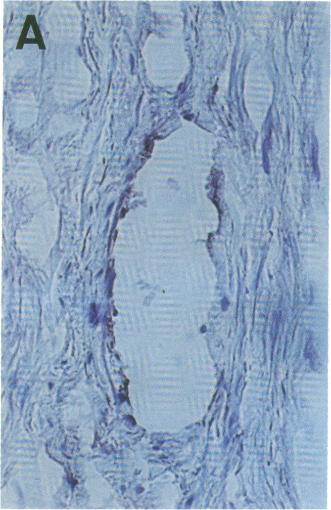
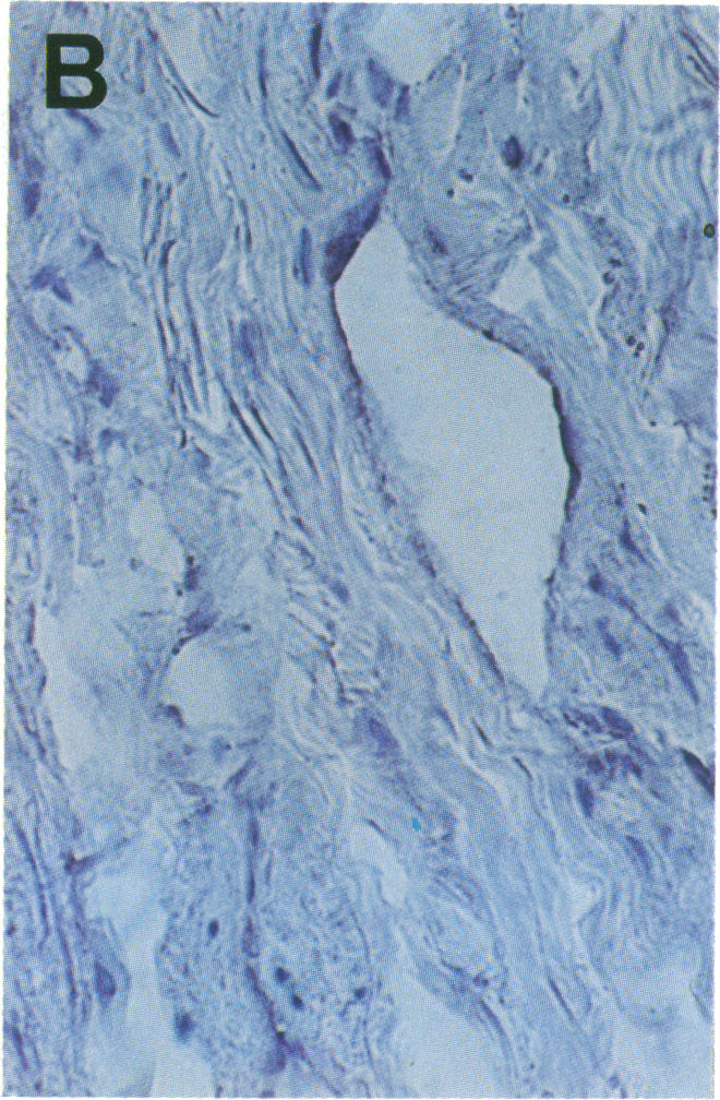
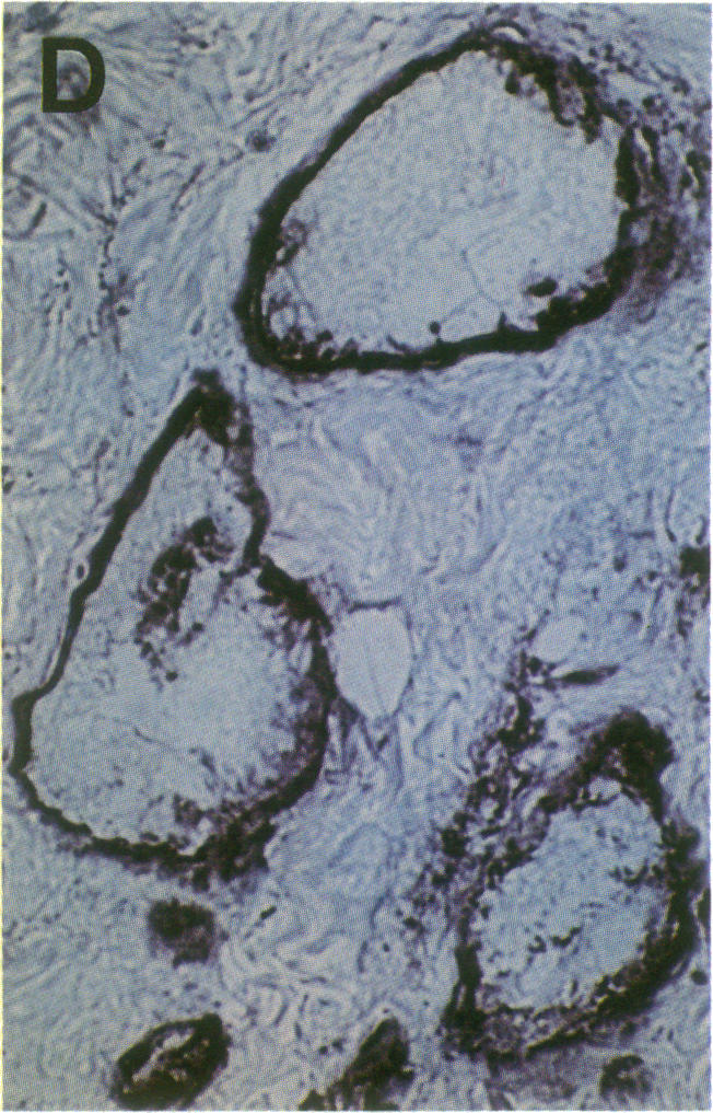
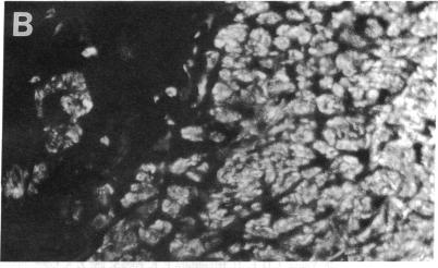
Cell-mediated autoimmunity has been strongly implicated in the pathogenesis of vascular cell injury in Takayasu's arteritis. To clarify the immunological mechanisms involved, we examined the expression of a cytolytic factor, perforin in infiltrating cells of aortic tissue samples from seven patients with Takayasu's arteritis. We also examined the expression of a 65-kD heat-shock protein (HSP-65), human leukocyte antigen classes I and II, and intercellular adhesion molecule-1 in the aortic tissue. Immunohistochemical studies showed that the infiltrating cells mainly consisted of gamma delta T lymphocytes, natural killer cells, macrophages, cytotoxic T lymphocytes and T helper cells, and that perforin was expressed in gamma delta T lymphocytes, natural killer cells, and cytotoxic T lymphocytes. In situ hybridization analysis also revealed expression of perforin mRNA in the infiltrating cells. Immunoelectron microscopic studies demonstrated that the infiltrating cells released massive amounts of perforin directly onto the surface of arterial vascular cells. We also found that expression of HSP-65, human leukocyte antigen classes I and II, and intercellular adhesion molecule-1 was strongly induced in the aortic tissue and might facilitate the recognition, adhesion and cytotoxicity of the infiltrating killer lymphocytes. These findings provide the first direct evidence that the infiltrating cells in the aortic tissue mainly consist of killer cells, and strongly suggest that these killer cells, especially gamma delta T lymphocytes, may recognize HSP-65 and play a critical role in the vascular cell injury of Takayasu's arteritis by releasing perforin.
细胞介导的自身免疫在高安动脉炎的血管细胞损伤发病机制中具有重要作用。为阐明其中的免疫机制,我们检测了7例高安动脉炎患者主动脉组织样本浸润细胞中细胞溶解因子穿孔素的表达。我们还检测了主动脉组织中65-kD热休克蛋白(HSP-65)、人类白细胞抗原I类和II类以及细胞间黏附分子-1的表达。免疫组织化学研究显示,浸润细胞主要由γδT淋巴细胞、自然杀伤细胞、巨噬细胞、细胞毒性T淋巴细胞和辅助性T细胞组成,穿孔素在γδT淋巴细胞、自然杀伤细胞和细胞毒性T淋巴细胞中表达。原位杂交分析也显示浸润细胞中有穿孔素mRNA表达。免疫电子显微镜研究表明,浸润细胞直接向动脉血管细胞表面释放大量穿孔素。我们还发现,主动脉组织中HSP-65、人类白细胞抗原I类和II类以及细胞间黏附分子-1的表达被强烈诱导,这可能有助于浸润的杀伤淋巴细胞的识别、黏附和细胞毒性作用。这些发现首次提供了直接证据,表明主动脉组织中的浸润细胞主要由杀伤细胞组成,并强烈提示这些杀伤细胞,尤其是γδT淋巴细胞,可能识别HSP-65,并通过释放穿孔素在高安动脉炎的血管细胞损伤中起关键作用。