• 文献检索
  • 文档翻译
  • 深度研究
  • 学术资讯
  • Suppr Zotero 插件Zotero 插件
  • 邀请有礼
  • 套餐&价格
  • 历史记录
应用&插件
Suppr Zotero 插件Zotero 插件浏览器插件Mac 客户端Windows 客户端微信小程序
定价
高级版会员购买积分包购买API积分包
服务
文献检索文档翻译深度研究API 文档MCP 服务
关于我们
关于 Suppr公司介绍联系我们用户协议隐私条款
关注我们

Suppr 超能文献

核心技术专利:CN118964589B侵权必究
粤ICP备2023148730 号-1Suppr @ 2026

文献检索

告别复杂PubMed语法,用中文像聊天一样搜索,搜遍4000万医学文献。AI智能推荐,让科研检索更轻松。

立即免费搜索

文件翻译

保留排版,准确专业,支持PDF/Word/PPT等文件格式,支持 12+语言互译。

免费翻译文档

深度研究

AI帮你快速写综述,25分钟生成高质量综述,智能提取关键信息,辅助科研写作。

立即免费体验

在愈合过程中的大鼠屈肌腱粘连形成期间,炎性细胞在滑膜鞘和腱旁组织中的积聚。

The accumulation of inflammatory cells in synovial sheath and epitenon during adhesion formation in healing rat flexor tendons.

作者信息

Wojciak B, Crossan J F

机构信息

Cell Biology Department, Glasgow University, UK.

出版信息

Clin Exp Immunol. 1993 Jul;93(1):108-14. doi: 10.1111/j.1365-2249.1993.tb06505.x.

DOI:10.1111/j.1365-2249.1993.tb06505.x
PMID:8324895
原文链接:https://pmc.ncbi.nlm.nih.gov/articles/PMC1554749/
Abstract

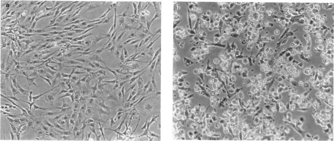
The accumulation of inflammatory cells in synovial tissue was studied using indirect immunofluorescence assays on cell cultures and frozen tissue sections of healing rat digital flexor tendons. Flexor tendons were collected from rats 3, 7 and 14 days after crush injury. Tendon sheath and epithenon cells were isolated by sequential enzymic digestion and cultured for 2 days. Subpopulations of synovial and inflammatory cells were identified with MoAbs against cell surface glycoproteins present on B lymphocytes (CD45), T lymphocytes (CD2, CD4, CD8), macrophages (CD14) and endothelial cells. A phagocytosis assay was also used to identify macrophages. We report a substantial increase in the number of T lymphocytes (mainly helper/inducer) and phagocytotic cells with monocyte/macrophage surface markers in tendon sheath and epitenon 3 days after crush injury. The infiltration of inflammatory cells into synovial sheath and epitenon preceded an increase in fibronectin production by tendon cells which was seen 7 days after injury. To study the interaction between T lymphocytes and synovial cells in vitro, we established synovial fibroblast-like type B cell cultures and used stimulated and non-stimulated T lymphocytes in cell binding assays. We observed increased adhesiveness between unstimulated synovial cells and synovial cells previously cultured with activated and non-activated T lymphocytes. ELISA inhibition studies have shown an increase in fibronectin production by synovial fibroblasts co-cultured with stimulated CD4+ T lymphocytes. We suggest that the presence of inflammatory cells in synovial sheath and epitenon during tendon healing induces synovial fibroblasts and epitenon cells to increase their production of fibronectin, which provides a scaffold for subsequent adhesion formation.

摘要

利用间接免疫荧光分析法,对愈合过程中的大鼠指屈肌腱的细胞培养物和冷冻组织切片进行研究,以观察滑膜组织中炎性细胞的聚集情况。在挤压伤后3天、7天和14天收集大鼠的屈肌腱。通过连续酶消化分离腱鞘和外皮细胞,并培养2天。用针对B淋巴细胞(CD45)、T淋巴细胞(CD2、CD4、CD8)、巨噬细胞(CD14)和内皮细胞表面存在的细胞表面糖蛋白的单克隆抗体来鉴定滑膜细胞和炎性细胞亚群。还采用吞噬试验来鉴定巨噬细胞。我们报告,挤压伤后3天,腱鞘和外皮中T淋巴细胞(主要是辅助/诱导性T淋巴细胞)以及具有单核细胞/巨噬细胞表面标志物的吞噬细胞数量大幅增加。炎性细胞浸润到滑膜鞘和外皮中,早于损伤后7天观察到的腱细胞纤连蛋白产生的增加。为了在体外研究T淋巴细胞与滑膜细胞之间的相互作用,我们建立了滑膜成纤维细胞样B细胞培养物,并在细胞结合试验中使用了经刺激和未经刺激的T淋巴细胞。我们观察到,未刺激的滑膜细胞与先前与活化和未活化T淋巴细胞共培养的滑膜细胞之间的黏附性增加。酶联免疫吸附测定抑制研究表明,与经刺激的CD4 + T淋巴细胞共培养的滑膜成纤维细胞产生的纤连蛋白增加。我们认为,肌腱愈合过程中滑膜鞘和外皮中炎性细胞的存在,诱导滑膜成纤维细胞和外皮细胞增加纤连蛋白的产生,这为随后的粘连形成提供了支架。

https://cdn.ncbi.nlm.nih.gov/pmc/blobs/07de/1554749/92cf79771c84/clinexpimmunol00032-0114-a.jpg
https://cdn.ncbi.nlm.nih.gov/pmc/blobs/07de/1554749/7ce962dce553/clinexpimmunol00032-0112-a.jpg
https://cdn.ncbi.nlm.nih.gov/pmc/blobs/07de/1554749/92cf79771c84/clinexpimmunol00032-0114-a.jpg
https://cdn.ncbi.nlm.nih.gov/pmc/blobs/07de/1554749/7ce962dce553/clinexpimmunol00032-0112-a.jpg
https://cdn.ncbi.nlm.nih.gov/pmc/blobs/07de/1554749/92cf79771c84/clinexpimmunol00032-0114-a.jpg

相似文献

1
The accumulation of inflammatory cells in synovial sheath and epitenon during adhesion formation in healing rat flexor tendons.在愈合过程中的大鼠屈肌腱粘连形成期间,炎性细胞在滑膜鞘和腱旁组织中的积聚。
Clin Exp Immunol. 1993 Jul;93(1):108-14. doi: 10.1111/j.1365-2249.1993.tb06505.x.
2
The effects of T cells and their products on in vitro healing of epitenon cell microwounds.T细胞及其产物对腱膜细胞微伤口体外愈合的影响。
Immunology. 1994 Sep;83(1):93-8.
3
Patterns of cellular activation after tendon injury.肌腱损伤后的细胞激活模式。
J Hand Surg Br. 1996 Dec;21(6):813-20. doi: 10.1016/s0266-7681(96)80199-9.
4
Flexor tendon wound healing in vitro: lactate up-regulation of TGF-beta expression and functional activity.体外屈肌腱伤口愈合:乳酸上调转化生长因子-β表达及功能活性
Plast Reconstr Surg. 2004 Feb;113(2):625-32. doi: 10.1097/01.PRS.0000101529.47062.34.
5
Flexor tendon wound healing in vitro: the effect of lactate on tendon cell proliferation and collagen production.体外屈肌腱伤口愈合:乳酸对肌腱细胞增殖和胶原蛋白产生的影响。
J Hand Surg Am. 2001 Sep;26(5):847-54. doi: 10.1053/jhsu.2001.26185.
6
Synovial sheath cell migratory response to flexor tendon injury: an experimental study in rats.滑膜鞘细胞对屈肌腱损伤的迁移反应:大鼠实验研究
J Hand Surg Am. 2003 Nov;28(6):987-93. doi: 10.1016/s0363-5023(03)00380-0.
7
Fibronectin in the tendon-synovial complex: quantitation in vivo and in vitro by ELISA and relative mRNA levels by polymerase chain reaction and northern blot.
J Orthop Res. 1994 Mar;12(2):253-61. doi: 10.1002/jor.1100120215.
8
In vitro flexor tendon cell response to TGF-beta1: a gene expression study.体外屈肌腱细胞对转化生长因子-β1的反应:一项基因表达研究。
J Hand Surg Am. 2009 Mar;34(3):495-503. doi: 10.1016/j.jhsa.2008.10.032.
9
The effect of constant direct electrical current on intrinsic healing in the flexor tendon in vitro. An ultrastructural study of differing attitudes in epitenon cells and tenocytes.恒定直流电对体外屈肌腱内在愈合的影响。腱外膜细胞和肌腱细胞不同状态的超微结构研究。
J Hand Surg Br. 1992 Feb;17(1):94-8. doi: 10.1016/0266-7681(92)90021-s.
10
Insulin-like growth factor-I is expressed by avian flexor tendon cells.
J Orthop Res. 2000 Jul;18(4):546-56. doi: 10.1002/jor.1100180406.

引用本文的文献

1
Challenges in tendon-bone healing: emphasizing inflammatory modulation mechanisms and treatment.肌腱-骨愈合的挑战:强调炎症调节机制和治疗。
Front Endocrinol (Lausanne). 2024 Nov 6;15:1485876. doi: 10.3389/fendo.2024.1485876. eCollection 2024.
2
Augmentation of Tendon and Ligament Repair with Fiber-Reinforced Hydrogel Composites.纤维增强水凝胶复合材料增强肌腱和韧带修复。
Adv Healthc Mater. 2024 Nov;13(29):e2400668. doi: 10.1002/adhm.202400668. Epub 2024 Aug 12.
3
The role of macrophage polarization in tendon healing and therapeutic strategies: Insights from animal models.

本文引用的文献

1
Digital flexor tendons: an experimental study. Part I. The significance of each component of the flexor mechanismhin tendon healing.指屈肌腱:一项实验研究。第一部分。屈肌机制各组成部分在肌腱愈合中的意义
Br J Plast Surg. 1960 Jan;12:289-316. doi: 10.1016/s0007-1226(59)80045-x.
2
THE FIBROBLAST IN FLEXOR TENDON HEALING.屈指肌腱愈合中的成纤维细胞。
Plast Reconstr Surg. 1964 Sep;34:223-32. doi: 10.1097/00006534-196409000-00001.
3
Tendon healing within the flexor digital sheath in the dog.
巨噬细胞极化在肌腱愈合中的作用及治疗策略:来自动物模型的见解
Front Bioeng Biotechnol. 2024 Feb 29;12:1366398. doi: 10.3389/fbioe.2024.1366398. eCollection 2024.
4
Effect of Aging on Tendon Biology, Biomechanics and Implications for Treatment Approaches.衰老对肌腱生物学、生物力学的影响及其对治疗方法的启示。
Int J Mol Sci. 2023 Oct 14;24(20):15183. doi: 10.3390/ijms242015183.
5
Mesenchymal stem cell licensing: enhancing MSC function as a translational approach for the treatment of tendon injury.间充质干细胞许可:增强 MSC 功能作为治疗肌腱损伤的转化方法。
Am J Vet Res. 2023 Sep 6;84(10):1-8. doi: 10.2460/ajvr.23.07.0154. Print 2023 Sep 1.
6
Innate and adaptive immune system cells implicated in tendon healing and disease.先天和适应性免疫系统细胞与肌腱愈合和疾病有关。
Eur Cell Mater. 2022 Feb 18;43:39-52. doi: 10.22203/eCM.v043a05.
7
Interplay of Forces and the Immune Response for Functional Tendon Regeneration.力的相互作用与功能性肌腱再生的免疫反应
Front Cell Dev Biol. 2021 Jun 4;9:657621. doi: 10.3389/fcell.2021.657621. eCollection 2021.
8
Local shifts in inflammatory and resolving lipid mediators in response to tendon overuse.局部炎症和解决脂类介质的变化对肌腱过度使用的反应。
FASEB J. 2021 Jun;35(6):e21655. doi: 10.1096/fj.202100078R.
9
The cellular basis of fibrotic tendon healing: challenges and opportunities.纤维性腱愈合的细胞基础:挑战与机遇。
Transl Res. 2019 Jul;209:156-168. doi: 10.1016/j.trsl.2019.02.002. Epub 2019 Feb 8.
10
Marked expression of TNF receptors in human peritendinous tissues including in nerve fascicles with axonal damage - Studies on tendinopathy and tennis elbow.肿瘤坏死因子受体在人类肌腱周围组织中显著表达,包括在伴有轴突损伤的神经束中——肌腱病和网球肘的研究。
J Musculoskelet Neuronal Interact. 2017 Sep 1;17(3):226-236.
J Bone Joint Surg Am. 1962 Jan;44-A:49-64.
4
Method to prove investigation of particles by macrophages with light microscopy.用光镜观察巨噬细胞对颗粒进行研究的方法。
Scand J Immunol. 1980;12(3):265-9. doi: 10.1111/j.1365-3083.1980.tb00066.x.
5
Superficial repair of severed flexor tendons in synovial environment. An experimental, ultrastructural study on cellular mechanisms.滑膜环境中屈指肌腱离断的浅层修复:细胞机制的实验性超微结构研究
J Hand Surg Am. 1980 Sep;5(5):451-61. doi: 10.1016/s0363-5023(80)80075-x.
6
The origin of type A synovial lining cells.A型滑膜衬里细胞的起源。
Immunobiology. 1982 Apr;161(3-4):227-31. doi: 10.1016/S0171-2985(82)80078-8.
7
The synovial cavity as a "tissue culture in situ"--science or nonsense?滑膜腔作为“原位组织培养”——科学还是无稽之谈?
J Hand Surg Am. 1982 Mar;7(2):196-9. doi: 10.1016/s0363-5023(82)80088-9.
8
Role of fibronectin in collagen deposition: Fab' to the gelatin-binding domain of fibronectin inhibits both fibronectin and collagen organization in fibroblast extracellular matrix.纤连蛋白在胶原蛋白沉积中的作用:针对纤连蛋白明胶结合结构域的Fab'抑制成纤维细胞外基质中纤连蛋白和胶原蛋白的组装。
J Cell Biol. 1982 Feb;92(2):485-92. doi: 10.1083/jcb.92.2.485.
9
Fibronectin is produced by blood vessels in response to injury.纤连蛋白是血管在受到损伤时产生的。
J Exp Med. 1982 Aug 1;156(2):646-51. doi: 10.1084/jem.156.2.646.
10
Flexor tendon healing and restoration of the gliding surface. An ultrastructural study in dogs.屈肌腱愈合与滑动面修复:犬的超微结构研究
J Bone Joint Surg Am. 1983 Jan;65(1):70-80.