MacKay S, Meyerhoff D J, Constans J M, Norman D, Fein G, Weiner M W
Magnetic Resonance Spectroscopy Unit, Department of Veterans Affairs Medical Center, San Francisco, USA.
Arch Neurol. 1996 Feb;53(2):167-74. doi: 10.1001/archneur.1996.00550020079018.
To use 1H magnetic resonance spectroscopic imaging to study differences in neuron density (N-acetylaspartate [NAA]), membrane phospholipid metabolites (choline [Cho]), and creatine-containing metabolites (creatine plus phosphocreatine [Cr]) in subjects with Alzheimer's disease (AD), with subcortical ischemic vascular dementia (SIVD), and elderly controls.
Cross-sectional, between groups.
A Veterans Affairs medical center and university memory clinic.
Forty elderly subjects with AD (n = 14), with SIVD (n = 8), and elderly controls (n = 18).
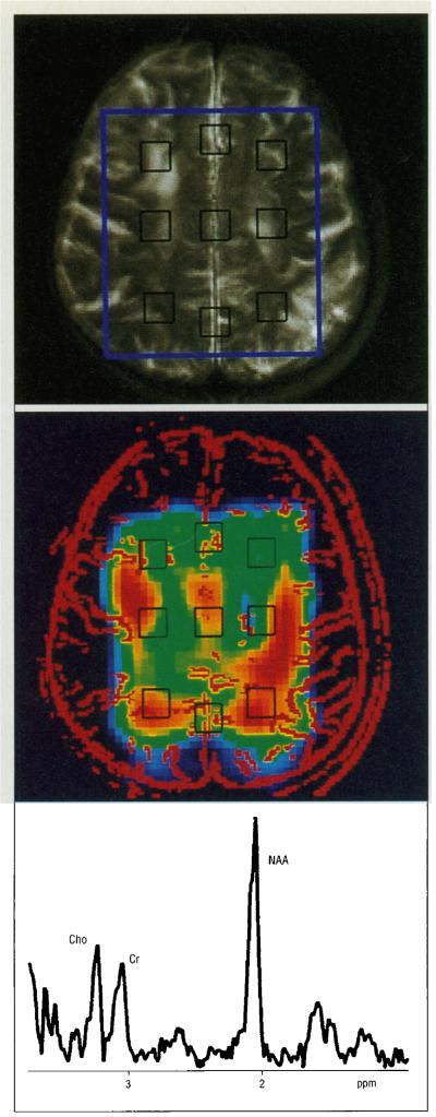
We used 1H magnetic resonance spectroscopic imaging to acquire spectra from a 80 x 100 x 17-mm volume superior to the lateral ventricles. Spectra were analyzed from voxels in anterior, medial, and posterior gray and white matter using nuclear magnetic resonance-1 and the results were compared between groups using repeated measures analysis of variance (ANOVA), Tukey's test, and individual Student's t tests.
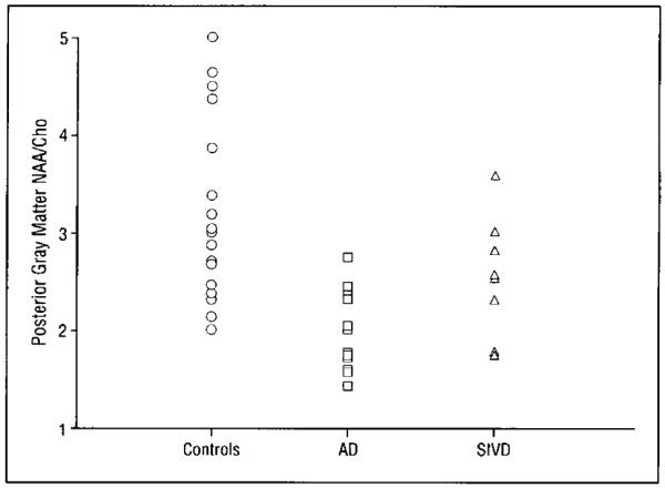
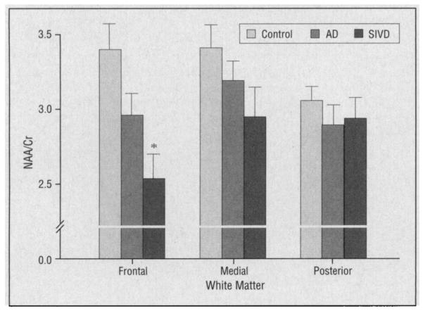
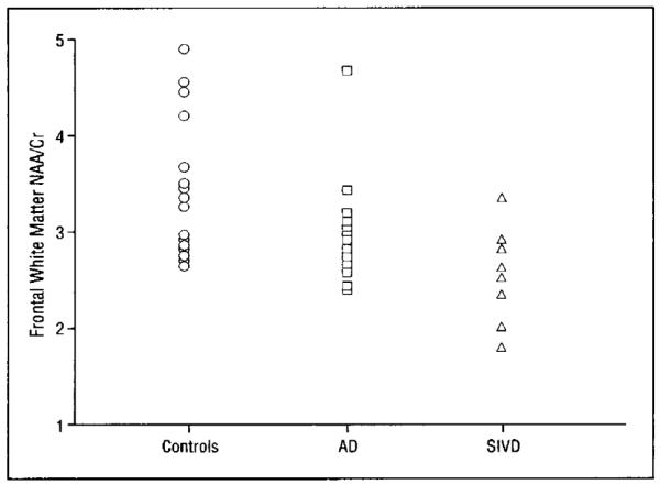
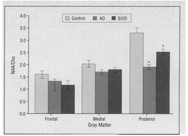
Using ANOVA, significantly lower levels of NAA/Cho and NAA/Cr and significantly higher levels of Cho/Cr were observed across both gray and white matter voxels in subjects with AD. Using individual Student's t tests, a significantly lower level of NAA/Cho and a higher level of Cho/Cr were observed in the posterior gray matter in subjects with AD. Using ANOVA in subjects with SIVD, significantly lower gray and white matter NAA/Cr levels were observed. Using Tukey's test, the NAA/Cr level was significantly lower in frontal white matter voxels in subjects with SIVD compared with controls.
Our findings in subjects with AD suggest neuron loss in gray matter, axon loss in white matter, and altered Cho metabolism in posterior brain regions. Our findings in subjects with SIVD are consistent with higher levels of creatine-containing metabolites and/or lower levels of NAA in frontal white matter.
使用氢质子磁共振波谱成像研究阿尔茨海默病(AD)患者、皮质下缺血性血管性痴呆(SIVD)患者及老年对照组在神经元密度(N - 乙酰天门冬氨酸 [NAA])、膜磷脂代谢物(胆碱 [Cho])和含肌酸代谢物(肌酸加磷酸肌酸 [Cr])方面的差异。
横断面研究,组间比较。
一家退伍军人事务医疗中心和大学记忆诊所。
40名老年受试者,其中AD患者14例,SIVD患者8例,老年对照组18例。
我们使用氢质子磁共振波谱成像从侧脑室上方一个80×100×17毫米的体积中获取波谱。使用核磁共振 - 1对前、中、后灰质和白质体素的波谱进行分析,并使用重复测量方差分析(ANOVA)、Tukey检验和个体学生t检验对组间结果进行比较。
使用ANOVA分析发现,AD患者的灰质和白质体素中NAA/Cho和NAA/Cr水平显著降低,Cho/Cr水平显著升高。使用个体学生t检验发现,AD患者后灰质中的NAA/Cho水平显著降低,Cho/Cr水平升高。对SIVD患者使用ANOVA分析发现,灰质和白质NAA/Cr水平显著降低。使用Tukey检验发现,与对照组相比,SIVD患者额叶白质体素中的NAA/Cr水平显著降低。
我们在AD患者中的研究结果表明灰质中存在神经元丢失、白质中存在轴突丢失以及后脑区域的Cho代谢改变。我们在SIVD患者中的研究结果与额叶白质中含肌酸代谢物水平较高和/或NAA水平较低一致。