Hofstad B, Vatn M H, Andersen S N, Huitfeldt H S, Rognum T, Larsen S, Osnes M
Medical Department, Ullevaal Hospital, Oslo, Norway.
Gut. 1996 Sep;39(3):449-56. doi: 10.1136/gut.39.3.449.
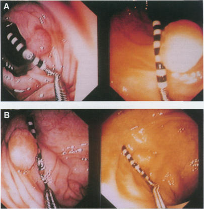
BACKGROUND, AIMS, AND PATIENTS: In a prospective follow up and intervention study of colorectal polyps, leaving all polyps less than 10 mm in situ for three years, analysis of redetection rate, growth, and new polyp formation was carried out in 116 patients undergoing annual colonoscopy. The findings in relation to growth and new polyp formation were applied to 58 subjects who received placebo.
Redetection rate varied from 75-90% for each year, and was highest in the rectum and sigmoid colon. There was no net change in size of all polyps in the placebo group, however, polyps less than 5 mm showed a tendency to net growth, and polyps 5-9 mm a tendency to net regression in size, both for adenomas and hyperplastic polyps. This pattern was verified by computerised image analysis. Patients between 50 and 60 years showed evidence of adenoma size increase compared with the older patients, and the same was true for those with multiple adenomas (four to five) compared with those with a single adenoma. The new adenomas were significantly smaller and 71% were located in the right side of the colon. Patients with multiple adenomas had more new polyps at all the follow up examinations than patients with a single adenoma. One patient developed an invasive colorectal carcinoma, which may be evolved from a previously overlooked polyp. Two polyps, showing intramucosal carcinoma after follow up for three years, were completely removed, as judged by endoscopy and histological examination.
The results show that follow up of unresected colorectal polyps up to 9 mm is safe. The consistency of growth retardation of medium sized polyps suggests extended intervals between the endoscopic follow up examinations, but the increased number of new polyps in the proximal colon indicates total colonoscopy as the examination of choice. The growth retardation of the medium sized polyps may partly explain the discrepancy between the prevalence of polyps and the incidence of colorectal cancer.
背景、目的和患者:在一项关于大肠息肉的前瞻性随访和干预研究中,对所有直径小于10毫米的息肉原位保留三年,对116例接受年度结肠镜检查的患者进行了再检出率、生长情况和新息肉形成情况的分析。将与生长和新息肉形成相关的研究结果应用于58例接受安慰剂治疗的受试者。
每年的再检出率在75%至90%之间,在直肠和乙状结肠中最高。安慰剂组所有息肉的大小没有净变化,然而,直径小于5毫米的息肉有净生长的趋势,直径5至9毫米的息肉无论是腺瘤还是增生性息肉都有大小净缩小的趋势。这一模式通过计算机图像分析得到了验证。50至60岁的患者与老年患者相比,腺瘤大小有增加的迹象,有多个腺瘤(四至五个)的患者与有单个腺瘤的患者相比也是如此。新腺瘤明显较小,71%位于结肠右侧。在所有随访检查中,有多个腺瘤的患者比有单个腺瘤的患者有更多新息肉。一名患者发生了浸润性结直肠癌,可能由先前被忽视的息肉演变而来。经内镜和组织学检查判断,两个息肉在随访三年后显示为黏膜内癌,已被完全切除。
结果表明,对直径达9毫米的未切除大肠息肉进行随访是安全的。中等大小息肉生长迟缓的一致性表明内镜随访检查的间隔时间可以延长,但近端结肠新息肉数量的增加表明全结肠镜检查是首选检查方法。中等大小息肉的生长迟缓可能部分解释了息肉患病率与结直肠癌发病率之间的差异。