Gaglio T, Dionne M A, Compton D A
Department of Biochemistry, Dartmouth Medical School, Hanover, New Hampshire 03755, USA.
J Cell Biol. 1997 Sep 8;138(5):1055-66. doi: 10.1083/jcb.138.5.1055.
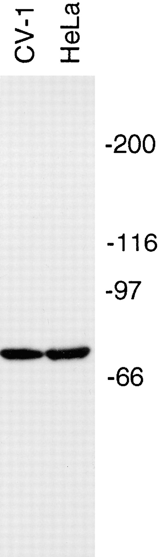
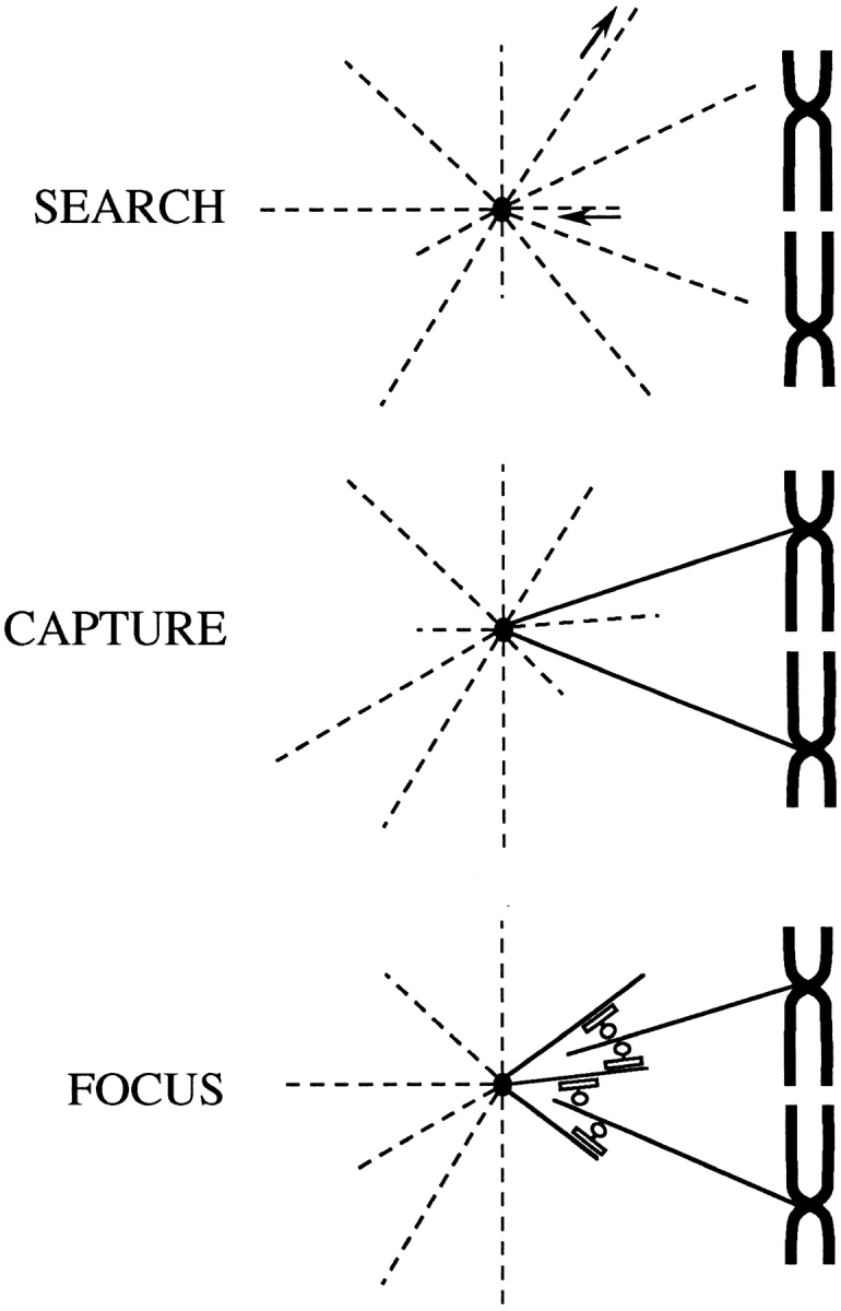
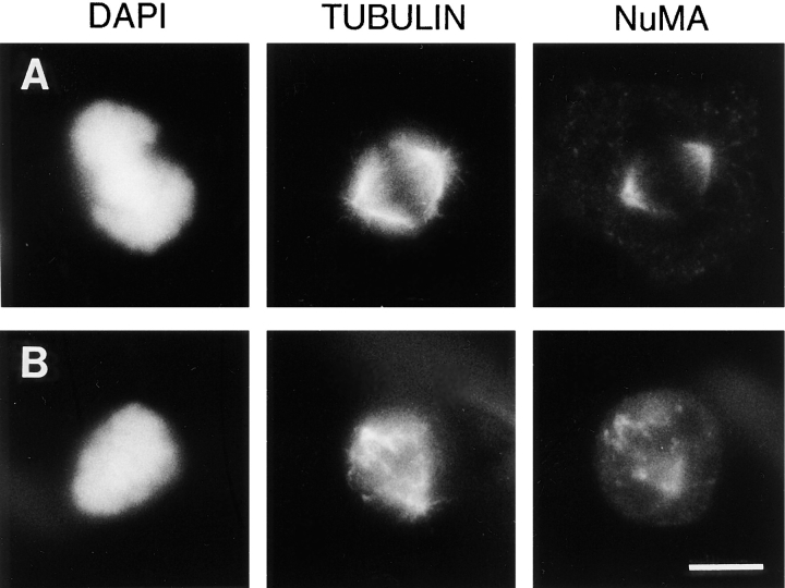
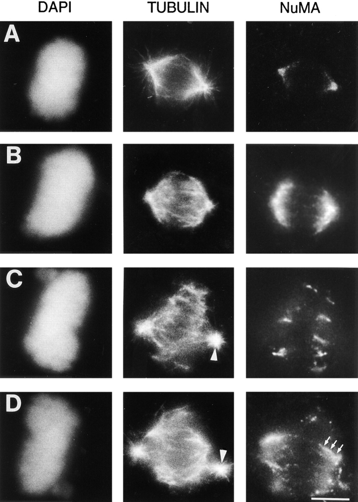
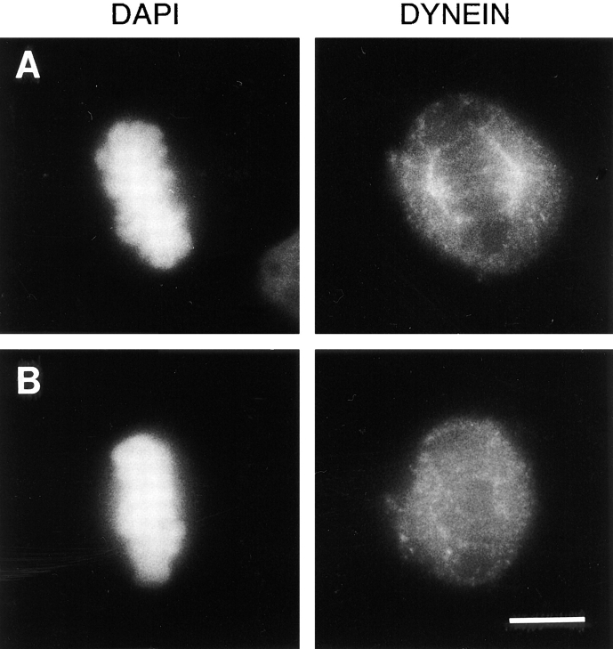
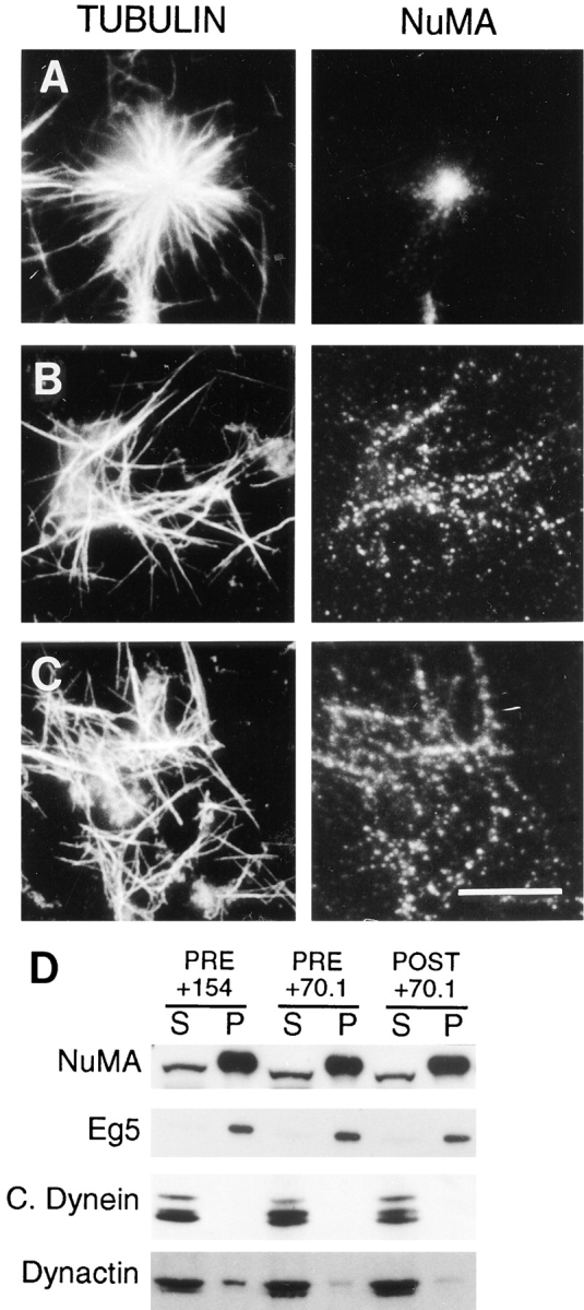
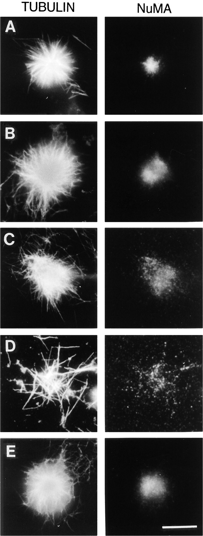
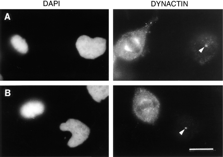
The focusing of microtubules into mitotic spindle poles in vertebrate somatic cells has been assumed to be the consequence of their nucleation from centrosomes. Contrary to this simple view, in this article we show that an antibody recognizing the light intermediate chain of cytoplasmic dynein (70.1) disrupts both the focused organization of microtubule minus ends and the localization of the nuclear mitotic apparatus protein at spindle poles when injected into cultured cells during metaphase, despite the presence of centrosomes. Examination of the effects of this dynein-specific antibody both in vitro using a cell-free system for mitotic aster assembly and in vivo after injection into cultured cells reveals that in addition to its direct effect on cytoplasmic dynein this antibody reduces the efficiency with which dynactin associates with microtubules, indicating that the antibody perturbs the cooperative binding of dynein and dynactin to microtubules during spindle/aster assembly. These results indicate that microtubule minus ends are focused into spindle poles in vertebrate somatic cells through a mechanism that involves contributions from both centrosomes and structural and microtubule motor proteins. Furthermore, these findings, together with the recent observation that cytoplasmic dynein is required for the formation and maintenance of acentrosomal spindle poles in extracts prepared from Xenopus eggs (Heald, R., R. Tournebize, T. Blank, R. Sandaltzopoulos, P. Becker, A. Hyman, and E. Karsenti. 1996. Nature (Lond.). 382: 420-425) demonstrate that there is a common mechanism for focusing free microtubule minus ends in both centrosomal and acentrosomal spindles. We discuss these observations in the context of a search-capture-focus model for spindle assembly.
脊椎动物体细胞中微管聚焦形成有丝分裂纺锤体极,一直被认为是微管从中心体成核的结果。与这种简单观点相反,在本文中我们表明,一种识别胞质动力蛋白轻链中间链的抗体(70.1),在中期注入培养细胞后,尽管存在中心体,但它会破坏微管负端的聚焦组织以及核有丝分裂器蛋白在纺锤体极的定位。在体外使用无细胞系统进行有丝分裂星状体组装以及在体内将抗体注入培养细胞后,对这种动力蛋白特异性抗体的作用进行检测发现,除了对胞质动力蛋白有直接作用外,该抗体还降低了动力蛋白激活蛋白与微管结合的效率,这表明该抗体在纺锤体/星状体组装过程中干扰了动力蛋白和动力蛋白激活蛋白与微管的协同结合。这些结果表明,脊椎动物体细胞中微管负端通过一种涉及中心体以及结构和微管运动蛋白共同作用的机制聚焦形成纺锤体极。此外,这些发现与最近的一项观察结果一致,即在非洲爪蟾卵提取物中,无中心体纺锤体极的形成和维持需要胞质动力蛋白(希尔德,R.,R. 图尔内比兹,T. 布兰克,R. 桑达尔佐普洛斯,P. 贝克尔,A. 海曼,和 E. 卡尔森蒂。1996. 《自然》(伦敦)。382: 420 - 425),这表明在有中心体和无中心体纺锤体中,聚焦游离微管负端存在一种共同机制。我们在纺锤体组装的搜索 - 捕获 - 聚焦模型的背景下讨论这些观察结果。