Centre for Bacterial Cell Biology, Institute for Cell and Molecular Biosciences, Newcastle University, Framlington Place, Newcastle upon Tyne, UK.
EMBO J. 2009 Aug 5;28(15):2272-82. doi: 10.1038/emboj.2009.129. Epub 2009 May 28.
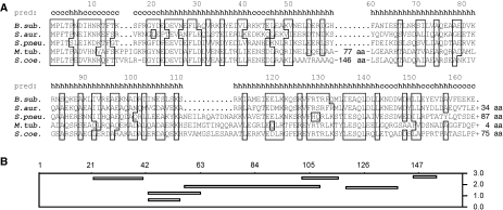
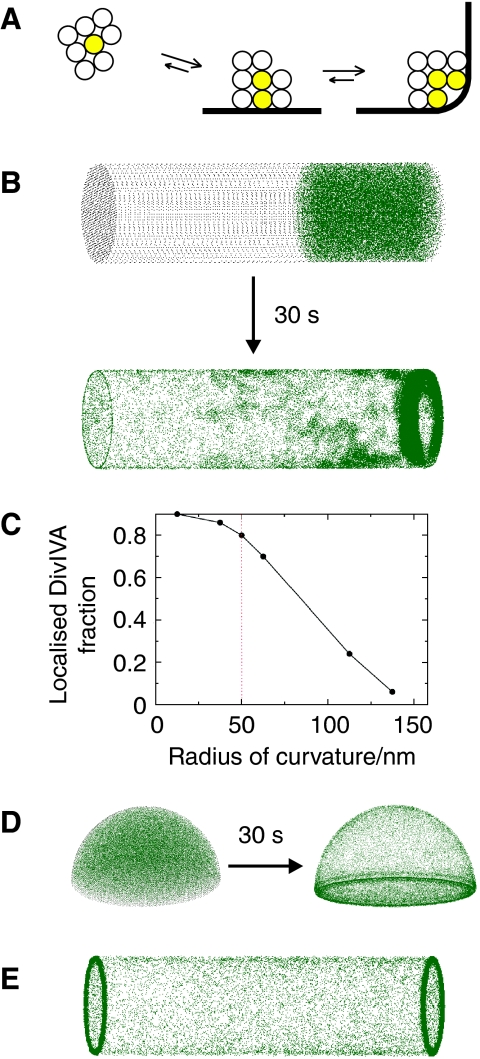
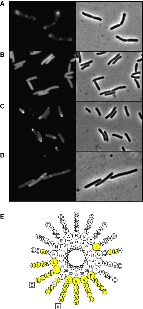
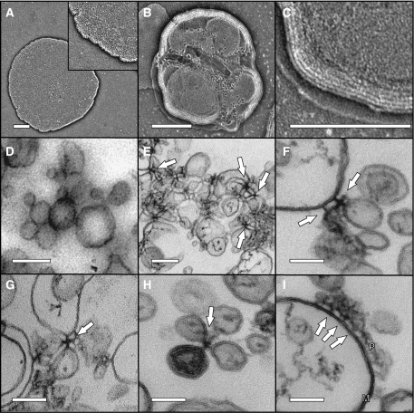
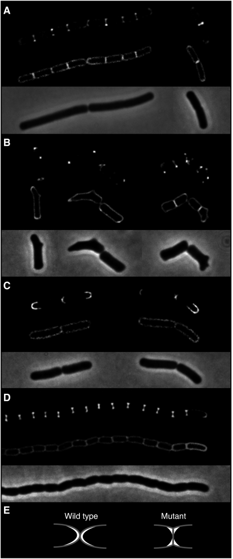
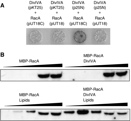
DivIVA is a conserved protein in Gram-positive bacteria and involved in various processes related to cell growth, cell division and spore formation. DivIVA is specifically targeted to cell division sites and cell poles. In Bacillus subtilis, DivIVA helps to localise other proteins, such as the conserved cell division inhibitor proteins, MinC/MinD, and the chromosome segregation protein, RacA. Little is known about the mechanism that localises DivIVA. Here we show that DivIVA binds to liposomes, and that the N terminus harbours the membrane targeting sequence. The purified protein can stimulate binding of RacA to membranes. In mutants with aberrant cell shapes, DivIVA accumulates where the cell membrane is most strongly curved. On the basis of electron microscopic studies and other data, we propose that this is due to molecular bridging of the curvature by DivIVA multimers. This model may explain why DivIVA localises at cell division sites. A Monte-Carlo simulation study showed that molecular bridging can be a general mechanism for binding of proteins to negatively curved membranes.
DivIVA 是革兰氏阳性菌中一种保守的蛋白质,参与与细胞生长、细胞分裂和孢子形成相关的各种过程。DivIVA 特异性靶向细胞分裂部位和细胞极。在枯草芽孢杆菌中,DivIVA 有助于定位其他蛋白质,如保守的细胞分裂抑制剂蛋白 MinC/MinD 和染色体分离蛋白 RacA。关于定位 DivIVA 的机制知之甚少。在这里,我们表明 DivIVA 与脂质体结合,并且 N 端含有膜靶向序列。纯化的蛋白质可以刺激 RacA 与膜的结合。在具有异常细胞形状的突变体中,DivIVA 在细胞膜弯曲最强的地方积累。基于电子显微镜研究和其他数据,我们提出这是由于 DivIVA 多聚体的分子桥接导致曲率。该模型可以解释为什么 DivIVA 定位在细胞分裂部位。蒙特卡罗模拟研究表明,分子桥接可能是蛋白质与负曲率膜结合的一般机制。