Department of Biochemistry and Biophysics, The Keck Center for Advanced Microscopy, University of California San Francisco, San Francisco, California, United States of America.
PLoS One. 2010 Sep 15;5(9):e12768. doi: 10.1371/journal.pone.0012768.
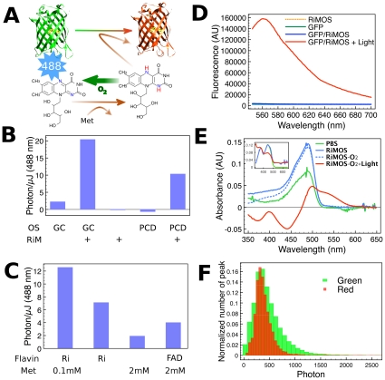
Photoactivated localization microscopy (PALM) and related fluorescent biological imaging methods are capable of providing very high spatial resolutions (up to 20 nm). Two major demands limit its widespread use on biological samples: requirements for photoactivatable/photoconvertible fluorescent molecules, which are sometimes difficult to incorporate, and high background signals from autofluorescence or fluorophores in adjacent focal planes in three-dimensional imaging which reduces PALM resolution significantly. We present here a high-resolution PALM method utilizing conventional EGFP as the photoconvertible fluorophore, improved algorithms to deal with high levels of biological background noise, and apply this to imaging higher order chromatin structure. We found that the emission wavelength of EGFP is efficiently converted from green to red when exposed to blue light in the presence of reduced riboflavin. The photon yield of red-converted EGFP using riboflavin is comparable to other bright photoconvertible fluorescent proteins that allow <20 nm resolution. We further found that image pre-processing using a combination of denoising and deconvolution of the raw PALM images substantially improved the spatial resolution of the reconstruction from noisy images. Performing PALM on Drosophila mitotic chromosomes labeled with H2AvD-EGFP, a histone H2A variant, revealed filamentous components of ∼70 nm. This is the first observation of fine chromatin filaments specific for one histone variant at a resolution approximating that of conventional electron microscope images (10-30 nm). As demonstrated by modeling and experiments on a challenging specimen, the techniques described here facilitate super-resolution fluorescent imaging with common biological samples.
光激活定位显微镜(PALM)和相关的荧光生物成像方法能够提供非常高的空间分辨率(高达 20nm)。有两个主要的需求限制了其在生物样本上的广泛应用:对可光激活/光转化的荧光分子的需求,有时难以掺入;以及在三维成像中来自自发荧光或相邻焦平面上荧光团的高背景信号,这大大降低了 PALM 的分辨率。我们在这里提出了一种利用传统 EGFP 作为光转化荧光团的高分辨率 PALM 方法,改进的算法来处理高水平的生物背景噪声,并将其应用于更高阶染色质结构的成像。我们发现,在存在还原核黄素的情况下,EGFP 的发射波长在暴露于蓝光时会有效地从绿色转换为红色。使用核黄素转化的红色 EGFP 的光子产率可与其他允许 <20nm 分辨率的亮敏光转化荧光蛋白相媲美。我们进一步发现,使用降噪和原始 PALM 图像的反卷积相结合的图像预处理,大大提高了从噪声图像重建的空间分辨率。用 H2AvD-EGFP 标记的果蝇有丝分裂染色体进行 PALM,H2A 变体,揭示了约 70nm 的丝状成分。这是首次观察到一种特定于一种组蛋白变体的精细染色质丝,分辨率接近传统电子显微镜图像(10-30nm)。通过对具有挑战性的样本进行建模和实验证明,这里描述的技术为具有常见生物样本的超分辨率荧光成像提供了便利。