Ocular Surface Center, Cullen Eye Institute, Department of Ophthalmology, Baylor College of Medicine, Houston, Texas 77030, USA.
Am J Ophthalmol. 2010 Dec;150(6):798-806. doi: 10.1016/j.ajo.2010.06.014.
To evaluate cross-sectional areas of conjunctivochalasis and tear meniscus using Fourier-Domain RTVue-100 optical coherence tomography (OCT) before and after conjunctival cauterization and to evaluate inter- and intraobserver reliability.
Prospective, nonrandomized, consecutive case study.
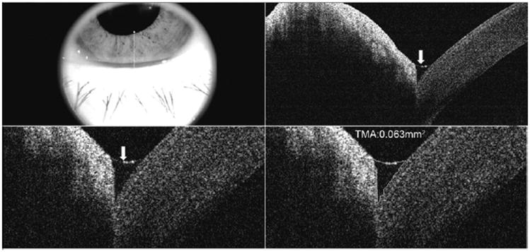
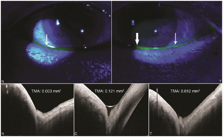
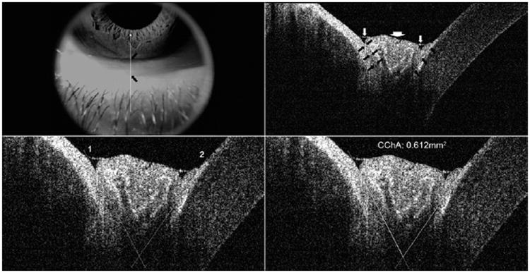
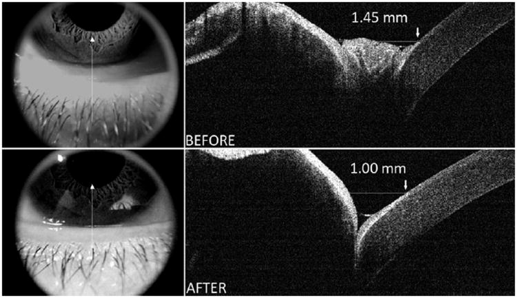
A total of 12 eyes of 7 patients with conjunctivochalasis (aged 56 to 87) were evaluated. After topical anesthesia, conjunctival cauterization was performed on the inferior bulbar conjunctiva. All patients underwent anterior segment OCT (AS-OCT) imaging prior to and 4 weeks after the procedure. Cross-sectional tear meniscus and conjunctivochalasis areas at 3 locations (nasal, center, and temporal areas) were measured in all patients.
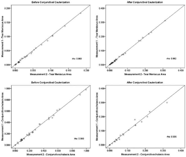
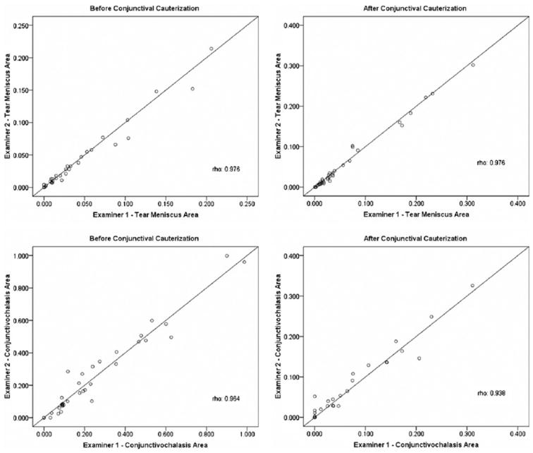
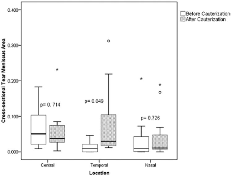
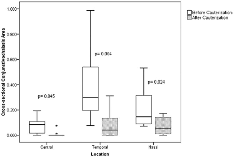
Nonsignificant increases (P = .177) in cross-sectional tear meniscus area as a whole (3 locations combined) were observed following cauterization. Cross-sectional conjunctivochalasis area measurements significantly decreased in all 3 locations after cauterization (P < .001). Mean cross-sectional conjunctivochalasis area decreased from 0.247 ± 0.24 mm(2) to 0.054 ± 0.79 mm(2). For 2 measurements of cross-sectional tear meniscus area by examiner 1, intraclass correlation coefficients ranged from 0.998 to 0.999. Among 2 examiners, Cronbach's alpha reliability coefficients were as high as 0.993 and 0.997 before and after conjunctival cauterization. Regarding the cross-sectional conjunctivochalasis area measurements, intraclass correlation coefficient values were similar to those of the cross-sectional tear meniscus area, but Cronbach's alpha reliability coefficients were slightly less.
This study indicates the AS-OCT is a useful and reproducible instrument to measure the cross-sectional area of conjunctiva prolapsing into the tear meniscus of patients with conjunctivochalasis. The method can monitor effectiveness of thermoreduction of conjunctivochalasis.
使用傅里叶域 RTVue-100 光学相干断层扫描(OCT)评估结膜松弛症患者结膜下凝固治疗前后的结膜穹隆横截面积和泪膜半月板,并评估观察者间和观察者内的可靠性。
前瞻性、非随机、连续病例研究。
本研究纳入了 7 例(56 至 87 岁)结膜松弛症患者的 12 只眼。局部麻醉后,在下穹隆结膜行结膜下凝固治疗。所有患者均在治疗前后进行眼前节 OCT(AS-OCT)成像。在所有患者中,测量了 3 个位置(鼻侧、中央和颞侧)的泪膜半月板和结膜松弛症的横截面积。
凝固治疗后,整个(3 个位置合并)泪膜半月板的横截面积无显著增加(P =.177)。凝固治疗后,所有 3 个位置的结膜松弛症横截面积均显著减小(P <.001)。平均结膜松弛症横截面积从 0.247 ± 0.24mm² 降至 0.054 ± 0.79mm²。对于观察者 1 的 2 次泪膜半月板横截面积测量,组内相关系数范围为 0.998 至 0.999。在 2 位观察者中,凝固治疗前后 Cronbach's alpha 可靠性系数高达 0.993 和 0.997。对于结膜松弛症的横截面积测量,组内相关系数值与泪膜半月板的横截面积相似,但 Cronbach's alpha 可靠性系数略低。
本研究表明 AS-OCT 是一种有用且可重复的仪器,可用于测量患有结膜松弛症患者的结膜突入泪膜半月板的横截面积。该方法可监测结膜松弛症热凝固治疗的效果。