Departments of Medicine and Laboratory Medicine, University of California, San Francisco, San Francisco, California, USA.
Nat Methods. 2011 Jan;8(1):91-6. doi: 10.1038/nmeth.1543. Epub 2010 Dec 12.
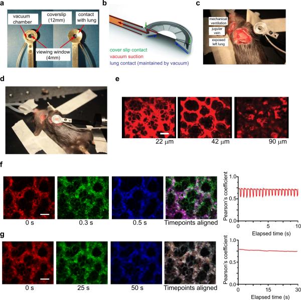
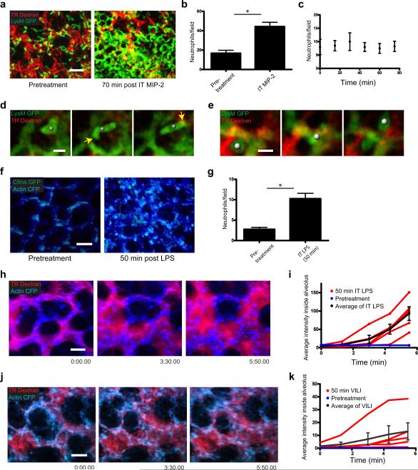
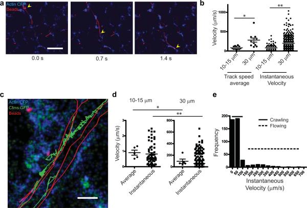
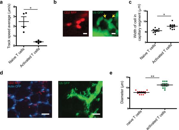
Real-time imaging of cellular and subcellular dynamics in vascularized organs requires image resolution and image registration to be simultaneously optimized without perturbing normal physiology. This problem is particularly pronounced in the lung, in which cells may transit at speeds >1 mm s(-1) and in which normal respiration results in large-scale tissue movements that prevent image registration. Here we report video-rate, two-photon imaging of a physiologically intact preparation of the mouse lung that is stabilizing and nondisruptive. Using our method, we obtained evidence for differential trapping of T cells and neutrophils in mouse pulmonary capillaries, and observed neutrophil mobilization and dynamic vascular leak in response to stretch and inflammatory models of lung injury in mice. The system permits physiological measurement of motility rates of >1 mm s(-1), observation of detailed cellular morphology and could be applied in the future to other organs and tissues while maintaining intact physiology.
在血管化器官中实时成像细胞和亚细胞动力学需要同时优化图像分辨率和图像配准,而不会干扰正常的生理机能。在肺部,这个问题尤为突出,细胞的迁移速度可能超过 1 毫米/秒,而正常呼吸会导致大规模的组织运动,从而无法进行图像配准。在这里,我们报告了一种视频速率的、对生理完整的小鼠肺部的双光子成像方法,该方法稳定且无损伤。使用我们的方法,我们获得了证据表明 T 细胞和中性粒细胞在小鼠肺毛细血管中的差异捕获,并且观察到中性粒细胞的动员以及对拉伸和炎症性肺损伤模型的动态血管渗漏。该系统可以实现超过 1 毫米/秒的运动速度的生理测量,观察详细的细胞形态,并且在未来可以应用于其他器官和组织,同时保持完整的生理机能。