Radiology and Imaging Sciences Department, National Institutes of Health Clinical Center, Building 10, Room 1C515, Bethesda, MD 20892-1182, USA.
Acad Radiol. 2012 May;19(5):562-70. doi: 10.1016/j.acra.2012.01.005. Epub 2012 Feb 15.
The aims of this study were to develop and validate an automated method to segment the renal cortex on contrast-enhanced abdominal computed tomographic images from kidney donors and to track cortex volume change after donation.
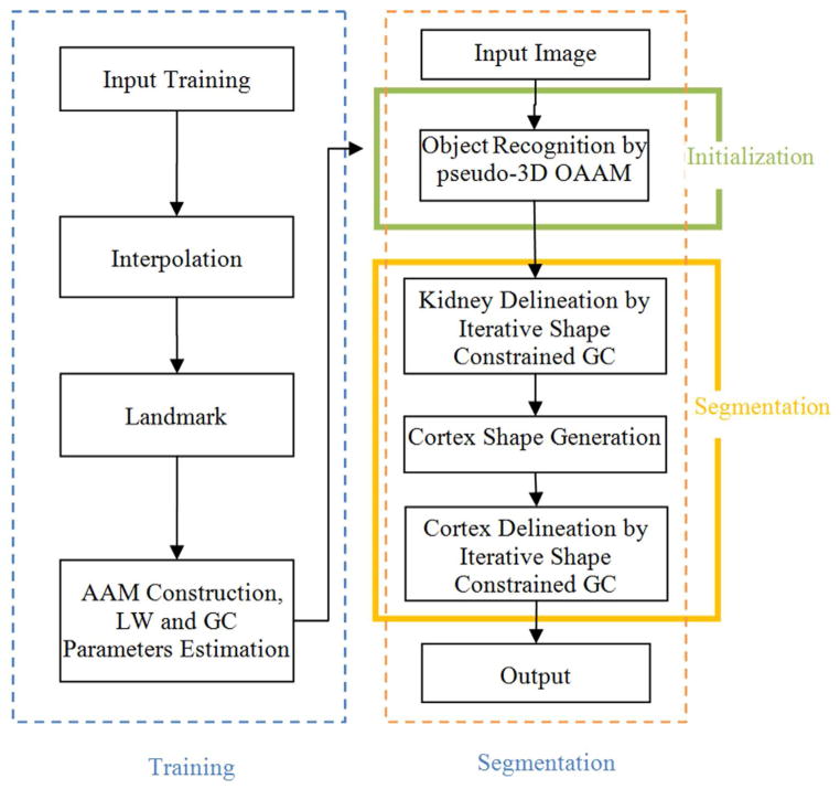
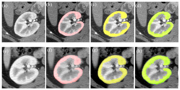
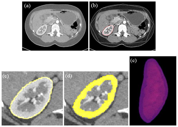
A three-dimensional fully automated renal cortex segmentation method was developed and validated on 37 arterial phase computed tomographic data sets (27 patients, 10 of whom underwent two computed tomographic scans before and after nephrectomy) using leave-one-out strategy. Two expert interpreters manually segmented the cortex slice by slice, and linear regression analysis and Bland-Altman plots were used to compare automated and manual segmentation. The true-positive and false-positive volume fractions were also calculated to evaluate the accuracy of the proposed method. Cortex volume changes in 10 subjects were also calculated.
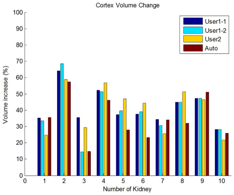
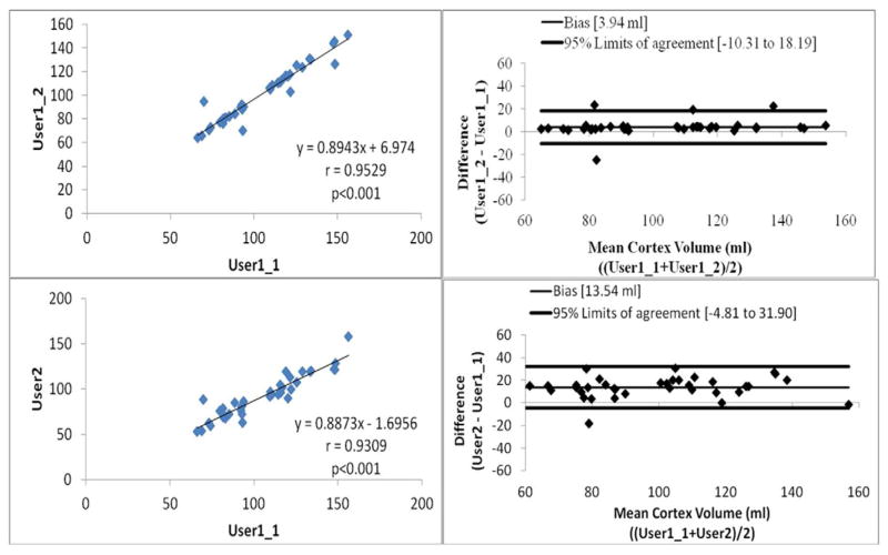
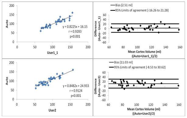
The linear regression analysis results showed that the automated and manual segmentation methods had strong correlations, with Pearson's correlations of 0.9529, 0.9309, 0.9283, and 0.9124 between intraobserver variation, interobserver variation, automated and user 1, and automated and user 2, respectively (P < .001 for all analyses). The Bland-Altman plots for cortex segmentation also showed that the automated and manual methods had agreeable segmentation. The mean volume increase of the cortex for the 10 subjects was 35.1 ± 13.2% (P < .01 by paired t test). The overall true-positive and false-positive volume fractions for cortex segmentation were 90.15 ± 3.11% and 0.85 ± 0.05%. With the proposed automated method, the time for cortex segmentation was reduced from 20 minutes for manual segmentation to 2 minutes.
The proposed method was accurate and efficient and can replace the current subjective and time-consuming manual procedure. The computer measurement confirms the volume of renal cortex increases after kidney donation.
本研究旨在开发和验证一种自动方法,以分割供体腹部增强 CT 图像的肾皮质,并跟踪捐献后的皮质体积变化。
使用留一法,在 37 个动脉期 CT 数据集(27 例患者,其中 10 例在肾切除术前和术后进行了两次 CT 扫描)上开发和验证了一种三维全自动肾皮质分割方法。两位专家口译员逐片手动分割皮质,并用线性回归分析和 Bland-Altman 图比较自动和手动分割。还计算了真阳性和假阳性体积分数,以评估所提出方法的准确性。还计算了 10 个受试者的皮质体积变化。
线性回归分析结果表明,自动和手动分割方法具有很强的相关性,观察者内变异、观察者间变异、自动和用户 1、自动和用户 2 之间的 Pearson 相关系数分别为 0.9529、0.9309、0.9283 和 0.9124(所有分析 P<0.001)。皮质分割的 Bland-Altman 图也表明,自动和手动方法具有可接受的分割。10 个受试者皮质体积的平均增加量为 35.1±13.2%(配对 t 检验 P<0.01)。皮质分割的总体真阳性和假阳性体积分数分别为 90.15±3.11%和 0.85±0.05%。使用所提出的自动方法,皮质分割的时间从手动分割的 20 分钟减少到 2 分钟。
所提出的方法准确高效,可以替代当前主观且耗时的手动过程。计算机测量证实肾脏捐献后肾皮质体积增加。