• 文献检索
  • 文档翻译
  • 深度研究
  • 学术资讯
  • Suppr Zotero 插件Zotero 插件
  • 邀请有礼
  • 套餐&价格
  • 历史记录
应用&插件
Suppr Zotero 插件Zotero 插件浏览器插件Mac 客户端Windows 客户端微信小程序
定价
高级版会员购买积分包购买API积分包
服务
文献检索文档翻译深度研究API 文档MCP 服务
关于我们
关于 Suppr公司介绍联系我们用户协议隐私条款
关注我们

Suppr 超能文献

核心技术专利:CN118964589B侵权必究
粤ICP备2023148730 号-1Suppr @ 2026

文献检索

告别复杂PubMed语法,用中文像聊天一样搜索,搜遍4000万医学文献。AI智能推荐,让科研检索更轻松。

立即免费搜索

文件翻译

保留排版,准确专业,支持PDF/Word/PPT等文件格式,支持 12+语言互译。

免费翻译文档

深度研究

AI帮你快速写综述,25分钟生成高质量综述,智能提取关键信息,辅助科研写作。

立即免费体验

在Descemet膜剥脱自动内皮角膜移植术中使用频域光学相干断层扫描技术。

Intraoperative use of spectral-domain optical coherence tomography during Descemet's stripping automated endothelial keratoplasty.

作者信息

Sng Chelvin Ca, Luengo Gimeno Federico, Mehta Jodhbir S, Htoon Hla Myint, Tan Donald T

机构信息

Singapore Eye Research Institute and Singapore National Eye Center, Singapore.

出版信息

Clin Ophthalmol. 2012;6:479-86. doi: 10.2147/OPTH.S28971. Epub 2012 Mar 23.

DOI:10.2147/OPTH.S28971
PMID:22536040
原文链接:https://pmc.ncbi.nlm.nih.gov/articles/PMC3334228/
Abstract

PURPOSE

To evaluate the intraoperative changes in the donor lenticule, recipient cornea, and the reduction of interface fluid thickness during Descemet's stripping and automated endothelial keratoplasty with EndoGlide™ (Angiotech Pharmaceuticals Inc, Vancouver, Canada) donor insertion, using intraoperative spectral-domain optical coherence tomography.

METHODS

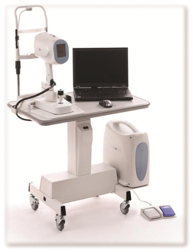
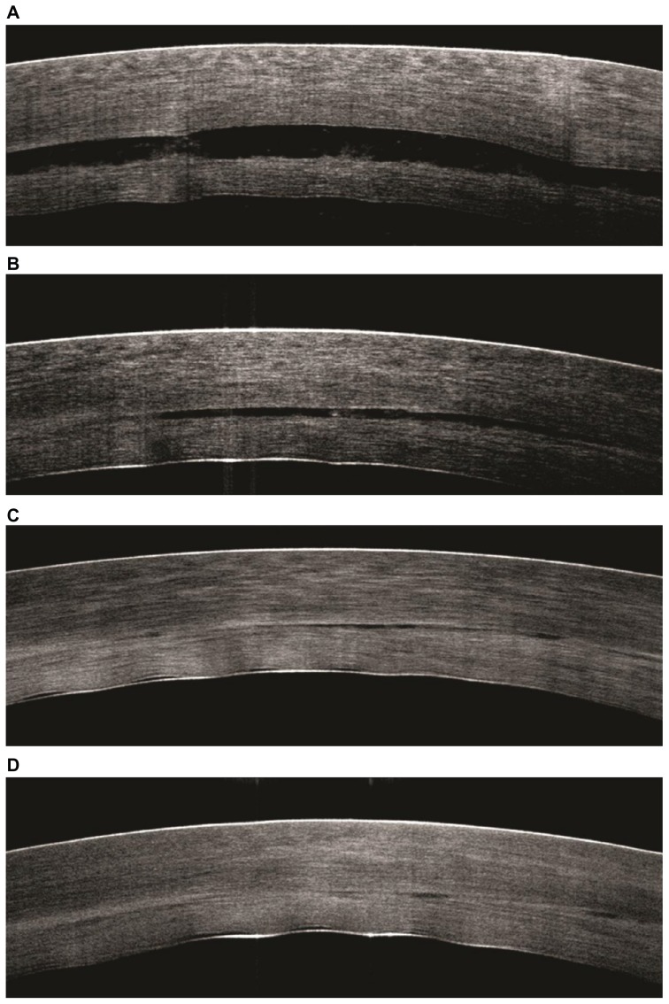
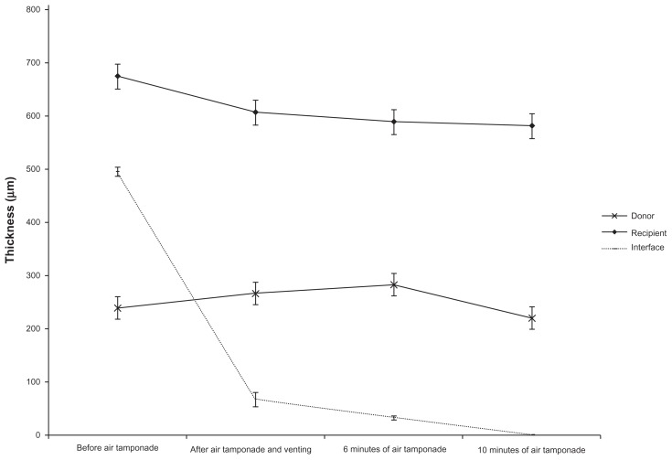
Prospective observational case series of patients underwent Descemet's stripping and automated endothelial keratoplasty using the EndoGlide inserter. Spectral-domain optical coherence tomography (iVue; Optovue Inc, Fremont, CA) with a handheld probe was used to image the cornea and anterior chamber. Standardized software was used to measure interface fluid gap, host cornea, and donor lenticule thicknesses during the following surgical stages of Descemet's stripping and automated endothelial keratoplasty: (1) after donor insertion and immediately before full air tamponade; (2) after air tamponade and expression of fluid from venting incisions; (3) at 6 minutes of air tamponade; and (4) at 10 minutes of air tamponade.

RESULTS

Ten patients with a mean age of 74.9 ± 11.8 years were recruited. Spectral-domain optical coherence tomography measurements of the interface fluid gap after fluid was expressed through the venting incisions (P < 0.001), at 6 minutes of air tamponade (P < 0.001) and at 10 minutes of air tamponade (P < 0.001 and P = 0.001, respectively), were significantly decreased compared to the measurements immediately before air tamponade. Donor thickness increased significantly at 6 minutes of air tamponade (P = 0.004) but reduced by 10 minutes compared to immediately before air tamponade.

CONCLUSION

Significant intraoperative changes in the donor, recipient cornea, and interface fluid thickness occurred following endothelial keratoplasty donor insertion.

摘要

目的

使用术中光谱域光学相干断层扫描技术,评估在Descemet膜剥离联合EndoGlide™(加拿大温哥华安格泰科制药公司)自动内皮角膜移植术中供体透镜、受体角膜的术中变化以及界面液厚度的减少情况。

方法

对接受使用EndoGlide插入器进行Descemet膜剥离联合自动内皮角膜移植术的患者进行前瞻性观察病例系列研究。使用带有手持探头的光谱域光学相干断层扫描(iVue;美国加利福尼亚州弗里蒙特市Optovue公司)对角膜和前房进行成像。在Descemet膜剥离联合自动内皮角膜移植术的以下手术阶段,使用标准化软件测量界面液间隙、宿主角膜和供体透镜厚度:(1)供体植入后且即将进行全空气填塞前;(2)空气填塞后且从排气切口排出液体后;(3)空气填塞6分钟时;(4)空气填塞10分钟时。

结果

招募了10名平均年龄为74.9±11.8岁的患者。与空气填塞前即刻的测量值相比,通过排气切口排出液体后(P<0.001)、空气填塞6分钟时(P<0.001)以及空气填塞10分钟时(分别为P<0.001和P = 0.001)的界面液间隙的光谱域光学相干断层扫描测量值显著降低。供体厚度在空气填塞6分钟时显著增加(P = 0.004),但与空气填塞前即刻相比,10分钟时有所减少。

结论

内皮角膜移植术供体植入后,供体、受体角膜及界面液厚度发生了显著的术中变化。

https://cdn.ncbi.nlm.nih.gov/pmc/blobs/8f94/3334228/d4eb2828b7bb/opth-6-479f3.jpg
https://cdn.ncbi.nlm.nih.gov/pmc/blobs/8f94/3334228/d2ef439a2fb1/opth-6-479f1.jpg
https://cdn.ncbi.nlm.nih.gov/pmc/blobs/8f94/3334228/3423df709ada/opth-6-479f2.jpg
https://cdn.ncbi.nlm.nih.gov/pmc/blobs/8f94/3334228/d4eb2828b7bb/opth-6-479f3.jpg
https://cdn.ncbi.nlm.nih.gov/pmc/blobs/8f94/3334228/d2ef439a2fb1/opth-6-479f1.jpg
https://cdn.ncbi.nlm.nih.gov/pmc/blobs/8f94/3334228/3423df709ada/opth-6-479f2.jpg
https://cdn.ncbi.nlm.nih.gov/pmc/blobs/8f94/3334228/d4eb2828b7bb/opth-6-479f3.jpg

相似文献

1
Intraoperative use of spectral-domain optical coherence tomography during Descemet's stripping automated endothelial keratoplasty.在Descemet膜剥脱自动内皮角膜移植术中使用频域光学相干断层扫描技术。
Clin Ophthalmol. 2012;6:479-86. doi: 10.2147/OPTH.S28971. Epub 2012 Mar 23.
2
Thin lenticule Descemet's stripping automated endothelial keratoplasty: single, slow pass technique.薄型角膜内皮切削术:单次、慢速撕囊技术。
Clin Exp Ophthalmol. 2014 Jul;42(5):411-6. doi: 10.1111/ceo.12271. Epub 2013 Dec 9.
3
Efficacy of Intraoperative Anterior Segment Optical Coherence Tomography during Descemet's Stripping Automated Endothelial Keratoplasty.在深板层角膜内皮移植术中术中前段光学相干断层扫描的疗效
ISRN Ophthalmol. 2014 Feb 2;2014:562062. doi: 10.1155/2014/562062. eCollection 2014.
4
Descemet's stripping endothelial automated keratoplasty using Tan EndoGlide endothelium insertion system.使用Tan EndoGlide内皮植入系统的Descemet膜剥除内皮自动角膜移植术
Transplant Proc. 2012 Nov;44(9):2759-64. doi: 10.1016/j.transproceed.2012.09.006.
5
Treatment of Descemet's membrane detachment after primary Descemet's stripping automated endothelial keratoplasty during surgery using intraoperative optical coherence tomography.术中光学相干断层扫描在原发性后弹力层剥除自动内皮角膜移植术手术中治疗后弹力层脱离
Am J Ophthalmol Case Rep. 2022 Jun 18;27:101623. doi: 10.1016/j.ajoc.2022.101623. eCollection 2022 Sep.
6
Visualization of precut DSAEK and pre-stripped DMEK donor corneas by intraoperative optical coherence tomography using the RESCAN 700.使用RESCAN 700通过术中光学相干断层扫描对预切割的深板层角膜内皮移植术(DSAEK)和预剥离的角膜后弹力层内皮移植术(DMEK)供体角膜进行可视化。
BMC Ophthalmol. 2016 Aug 4;16:135. doi: 10.1186/s12886-016-0308-z.
7
Histopathologic and optical coherence tomography observation after descemet's stripping endothelial keratoplasty in rabbits.兔后弹力层剥除内皮角膜移植术后的组织病理学及光学相干断层扫描观察
Exp Clin Transplant. 2014 Dec;12(6):548-54.
8
Outcomes of rebubbling for graft detachment after Descemet's stripping endothelial keratoplasty or Descemet's stripping automated endothelial keratoplasty.后撕囊内皮角膜移植或撕囊自动化内皮角膜移植后复灌处理植片脱离的结果。
Indian J Ophthalmol. 2020 Jan;68(1):48-53. doi: 10.4103/ijo.IJO_1521_18.
9
Theoretical, experimental, and optical coherence tomography (OCT) studies of graft apposition and adhesion in Descemets stripping automated endothelial keratoplasty (DSAEK).Descemets 撕囊全自动角膜内皮移植术中移植物贴附和黏附的理论、实验和光学相干断层扫描(OCT)研究。
Invest Ophthalmol Vis Sci. 2012 Jun 20;53(7):3839-46. doi: 10.1167/iovs.12-9593.
10
Optimizing outcomes with Descemet's membrane endothelial keratoplasty.优化 Descemet 膜内皮角膜移植术的结果。
Curr Opin Ophthalmol. 2014 Jul;25(4):325-34. doi: 10.1097/ICU.0000000000000072.

引用本文的文献

1
The Development and Clinical Application of Innovative Optical Ophthalmic Imaging Techniques.创新光学眼科成像技术的发展与临床应用
Front Med (Lausanne). 2022 Jun 30;9:891369. doi: 10.3389/fmed.2022.891369. eCollection 2022.
2
Clinical applications for intraoperative optical coherence tomography: a systematic review.术中光学相干断层扫描的临床应用:系统评价。
Eye (Lond). 2022 Feb;36(2):379-391. doi: 10.1038/s41433-021-01686-9. Epub 2021 Jul 16.
3
Intraoperative optical coherence tomography guided corneal sweeping for removal of remnant Interface fluid during ultra-thin Descemet stripping automated endothelial keratoplasty.

本文引用的文献

1
Quantitative assessment of changes in trabeculectomy blebs after laser suture lysis using anterior segment coherence tomography.应用眼前节相干断层扫描仪定量评估激光松解缝线后小梁切除术滤过泡的变化。
J Glaucoma. 2012 Jun-Jul;21(5):313-7. doi: 10.1097/IJG.0b013e31820e2d23.
2
Changes in retinal nerve fibre layer, optic nerve head morphology, and visual field after acute primary angle closure.急性原发性闭角型青光眼后视网膜神经纤维层、视盘形态和视野的变化。
Eye (Lond). 2011 May;25(5):619-25. doi: 10.1038/eye.2011.31. Epub 2011 Mar 25.
3
Laser in situ keratomileusis flap measurements: Comparison between observers and between spectral-domain and time-domain anterior segment optical coherence tomography.
术中光学相干断层扫描引导角膜清扫术在超薄Descemet膜剥除自动内皮角膜移植术中用于清除残余界面液。
BMC Ophthalmol. 2021 Apr 15;21(1):180. doi: 10.1186/s12886-021-01934-2.
4
Descemet membrane endothelial keratoplasty: intraoperative and postoperative imaging spectral-domain optical coherence tomography.Descemet膜内皮角膜移植术:术中及术后成像的频域光学相干断层扫描
Case Rep Ophthalmol Med. 2015;2015:506251. doi: 10.1155/2015/506251. Epub 2015 Feb 26.
激光原位角膜磨镶术瓣测量:观察者之间以及频域和时域眼前节光学相干断层扫描之间的比较。
J Cataract Refract Surg. 2011 Mar;37(3):544-51. doi: 10.1016/j.jcrs.2010.10.037.
4
Central corneal thickness measurements with Fourier-domain optical coherence tomography versus ultrasonic pachymetry and rotating Scheimpflug camera.傅里叶域光学相干断层扫描与超声角膜测厚仪和旋转式共焦激光角膜显微镜测量中央角膜厚度的比较。
Cornea. 2011 Jun;30(6):615-9. doi: 10.1097/ICO.0b013e3181d00800.
5
Descemet stripping automated endothelial keratoplasty with a graft insertion device: surgical technique and early clinical results.撕囊自动化角膜内皮移植术联合移植片植入装置:手术技术与早期临床结果。
Am J Ophthalmol. 2011 Feb;151(2):223-32.e2. doi: 10.1016/j.ajo.2010.08.027. Epub 2010 Dec 18.
6
Epithelial ingrowth after Descemet stripping automated endothelial keratoplasty: description of cases and assessment with anterior segment optical coherence tomography.Descemet 膜撕除自动化内皮角膜移植术后上皮内长入:病例描述和前节光学相干断层扫描评估。
Cornea. 2011 May;30(5):528-34. doi: 10.1097/ICO.0b013e3181fb8149.
7
Use of intraoperative fourier-domain anterior segment optical coherence tomography during descemet stripping endothelial keratoplasty.术中傅里叶域眼前节光学相干断层扫描在撕囊式内皮角膜移植术中的应用。
Am J Ophthalmol. 2010 Sep;150(3):360-365.e2. doi: 10.1016/j.ajo.2010.04.017. Epub 2010 Jun 29.
8
Pachymetric mapping with Fourier-domain optical coherence tomography.应用傅里叶域光学相干断层成像技术进行角膜生物力学测量。
J Cataract Refract Surg. 2010 May;36(5):826-31. doi: 10.1016/j.jcrs.2009.11.016.
9
Intra-examiner repeatability and agreement of corneal pachymetry map measurement by time-domain and Fourier-domain optical coherence tomography.时域和傅里叶域光学相干断层扫描测量角膜生物力学参数图的观察者内重复性和一致性。
Graefes Arch Clin Exp Ophthalmol. 2010 Nov;248(11):1647-56. doi: 10.1007/s00417-010-1360-7. Epub 2010 Mar 30.
10
Intraoperative use of three-dimensional spectral-domain optical coherence tomography.术中使用三维光谱域光学相干断层扫描技术。
Ophthalmic Surg Lasers Imaging. 2010 Mar-Apr;41(2):250-4. doi: 10.3928/15428877-20100303-15.