Wang Tianyi, Halaney David, Ho Derek, Feldman Marc D, Milner Thomas E
Department of Biomedical Engineering, University of Texas at Austin, 1 University Station C0800, Austin, Texas 78712, USA.
Biomed Opt Express. 2013 Apr 1;4(4):584-95. doi: 10.1364/BOE.4.000584. Epub 2013 Mar 21.
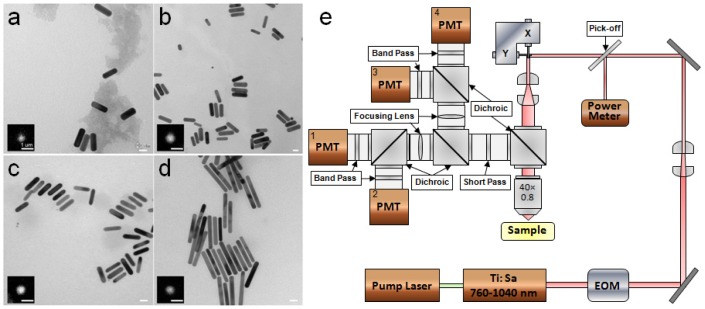
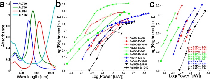
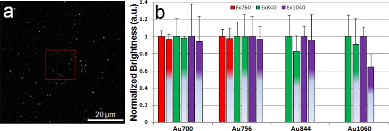
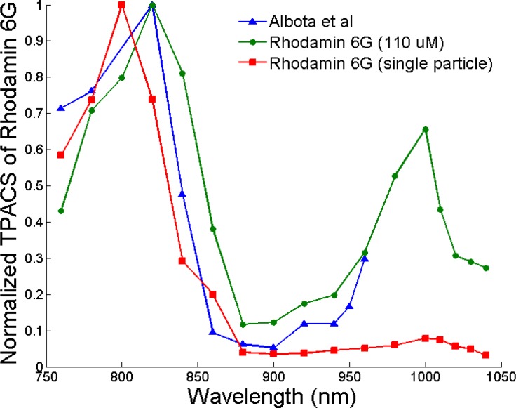
Gold nanorods can be internalized by macrophages (an important early cellular marker in atherosclerosis and cancer) and used as an imaging contrast agent for macrophage targeting. Objective of this study is to compare two-photon luminescence (TPL) properties of four aspect ratios of gold nanorods with surface plasmon resonance at 700, 756, 844 and 1060 nm respectively. TPL from single nanorods and Rhodamine 6G particles was measured using a laser-scanning TPL microscope. Nanorod TPL emission spectrum was recorded by a spectrometer. Quadratic dependence of luminescence intensity on excitation power (confirming a TPL process) was observed below a threshold (e.g., <1.6 mW), followed by photobleaching at higher power levels. Dependence of nanorod TPL intensity on excitation wavelength indicated that the two-photon action cross section (TPACS) is plasmon-enhanced. Largest TPACS of a single nanorod (12271 GM) was substantially larger than a single Rhodamine 6G particle (25 GM) at 760 nm excitation. Characteristics of nanorod TPL emission spectrum can be explained by plasmon-enhanced interband transition of gold. Comparison results of TPL brightness, TPACS and emission spectrum of nanorods can guide selection of optimal contrast agent for selected imaging applications.
金纳米棒可被巨噬细胞(动脉粥样硬化和癌症中一种重要的早期细胞标志物)内化,并用作巨噬细胞靶向的成像造影剂。本研究的目的是比较四种纵横比的金纳米棒在700、756、844和1060 nm处的表面等离子体共振与双光子发光(TPL)特性。使用激光扫描TPL显微镜测量单个纳米棒和罗丹明6G颗粒的TPL。用光谱仪记录纳米棒TPL发射光谱。在阈值以下(例如,<1.6 mW)观察到发光强度对激发功率的二次依赖性(证实了TPL过程),随后在较高功率水平下发生光漂白。纳米棒TPL强度对激发波长的依赖性表明双光子作用截面(TPACS)是等离子体增强的。在760 nm激发下,单个纳米棒的最大TPACS(12271 GM)明显大于单个罗丹明6G颗粒(25 GM)。纳米棒TPL发射光谱的特征可以通过金的等离子体增强带间跃迁来解释。纳米棒的TPL亮度、TPACS和发射光谱的比较结果可以指导为选定的成像应用选择最佳造影剂。