Nishimura Kei, Hiramatsu Kohzy, Monir Mohammad M, Takemoto Chihiro, Watanabe Takafumi
Department of Food Production Science, Graduate School of Agriculture, Shinshu University, 8304 Minami-minowa, Kami-ina, Nagano 399-4598, Japan.
J Vet Med Sci. 2013 Oct;75(10):1335-9. doi: 10.1292/jvms.13-0106. Epub 2013 Jul 12.
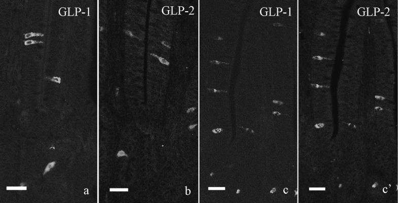
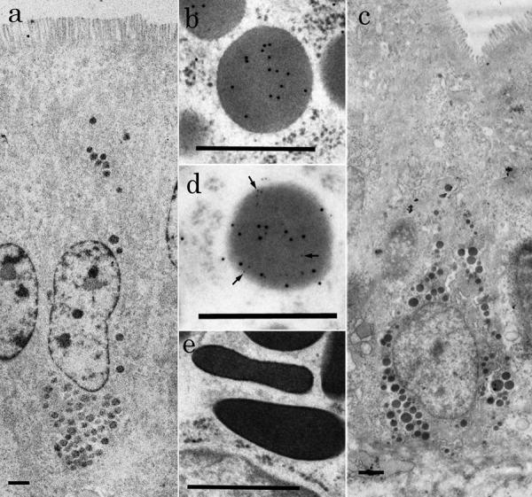
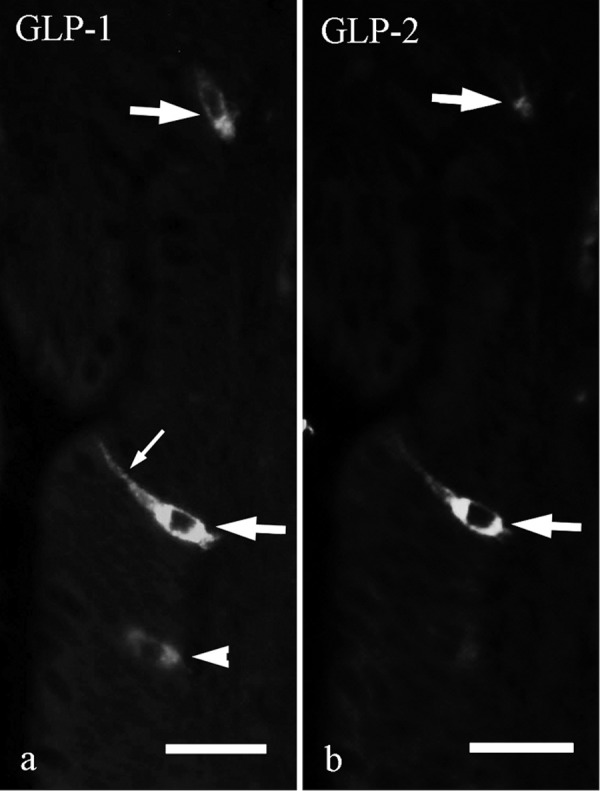
Colocalization of glucagon-like peptide (GLP)-1 with GLP-2 in L-cells was investigated in the chicken ileum by using double immunofluorescent and immunocytochemical techniques. Ultrastructural features of L-cells were also clarified in this study. L-cells showing immunoreactivity for both GLP-1 and GLP-2 were distributed in the whole ileum. They showed comma-like or flask-like shape and were located in epithelium of crypts and lower part of intestinal villi. L-cells showing GLP-1-immunoreactivity only were found in epithelium of lower and middle parts of intestinal villi. Transmission electron microscopy indicated that L-cells identified by colloidal gold-labeled immunocytochemistry were covered apically with microvilli, open-type and contained many secretory granules in their perikarya. These secretory granules without halo were round to oval in shape and showed moderate electron density. The longest and shortest diameters of secretory granules were 355 ± 62 nm (mean ± SD) and 287 ± 48 nm, respectively. Double labeling immunocytochemistry using two different sizes of particles (6 and 12 nm in diameter) of colloidal gold revealed that GLP-1 colocalized with GLP-2 in the same secretory granules. This study advances new morphological data about the endocrine system of the chicken small intestine.
采用双重免疫荧光和免疫细胞化学技术,在鸡回肠中研究了胰高血糖素样肽(GLP)-1与GLP-2在L细胞中的共定位情况。本研究还阐明了L细胞的超微结构特征。对GLP-1和GLP-2均呈免疫反应性的L细胞分布于整个回肠。它们呈逗号状或烧瓶状,位于隐窝上皮和肠绒毛下部。仅显示GLP-1免疫反应性的L细胞见于肠绒毛中下部的上皮。透射电子显微镜显示,经胶体金标记免疫细胞化学鉴定的L细胞顶端覆盖有微绒毛,为开放型,其核周质含有许多分泌颗粒。这些无晕的分泌颗粒呈圆形至椭圆形,电子密度适中。分泌颗粒的最长和最短直径分别为355±62nm(平均值±标准差)和287±48nm。使用两种不同大小(直径分别为6和12nm)的胶体金颗粒进行双重标记免疫细胞化学显示,GLP-1与GLP-2共定位于同一分泌颗粒中。本研究提供了关于鸡小肠内分泌系统的新形态学数据。