• 文献检索
  • 文档翻译
  • 深度研究
  • 学术资讯
  • Suppr Zotero 插件Zotero 插件
  • 邀请有礼
  • 套餐&价格
  • 历史记录
应用&插件
Suppr Zotero 插件Zotero 插件浏览器插件Mac 客户端Windows 客户端微信小程序
定价
高级版会员购买积分包购买API积分包
服务
文献检索文档翻译深度研究API 文档MCP 服务
关于我们
关于 Suppr公司介绍联系我们用户协议隐私条款
关注我们

Suppr 超能文献

核心技术专利:CN118964589B侵权必究
粤ICP备2023148730 号-1Suppr @ 2026

文献检索

告别复杂PubMed语法,用中文像聊天一样搜索,搜遍4000万医学文献。AI智能推荐,让科研检索更轻松。

立即免费搜索

文件翻译

保留排版,准确专业,支持PDF/Word/PPT等文件格式,支持 12+语言互译。

免费翻译文档

深度研究

AI帮你快速写综述,25分钟生成高质量综述,智能提取关键信息,辅助科研写作。

立即免费体验

应用二维差异凝胶电泳技术鉴定传染性胃肠炎冠状病毒感染 ST 细胞后的细胞蛋白质组。

Identification of cellular proteome using two-dimensional difference gel electrophoresis in ST cells infected with transmissible gastroenteritis coronavirus.

机构信息

Division of Swine Infectious Diseases, State Key Laboratory of Veterinary Biotechnology, Harbin Veterinary Research Institute, Chinese Academy of Agricultural Sciences, No, 427 Maduan Street, Nangang District, Harbin 150001, China.

出版信息

Proteome Sci. 2013 Jul 16;11(1):31. doi: 10.1186/1477-5956-11-31.

DOI:10.1186/1477-5956-11-31
PMID:23855489
原文链接:https://pmc.ncbi.nlm.nih.gov/articles/PMC3734006/
Abstract

BACKGROUND

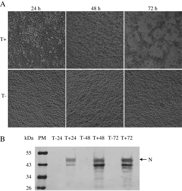
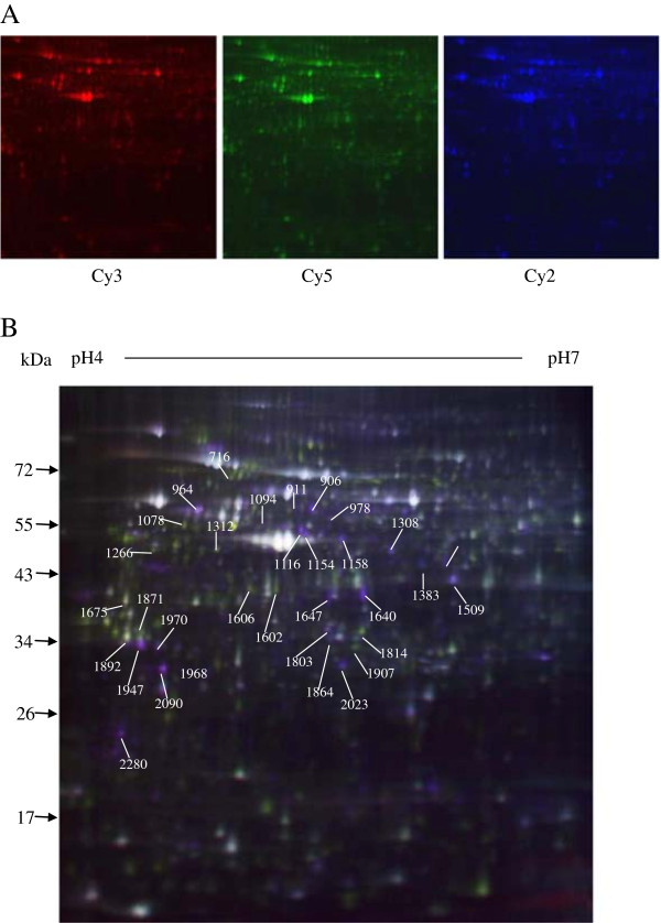
Transmissible gastroenteritis coronavirus (TGEV) is an enteropathogenic coronavirus that causes diarrhea in pigs, which is correlated with high morbidity and mortality in suckling piglets. Information remains limited about the comparative protein expression of host cells in response to TGEV infection. In this study, cellular protein response to TGEV infection in swine testes (ST) cells was analyzed, using the proteomic method of two-dimensional difference gel electrophoresis (2D DIGE) coupled with MALDI-TOF-TOF/MS identification.

RESULTS

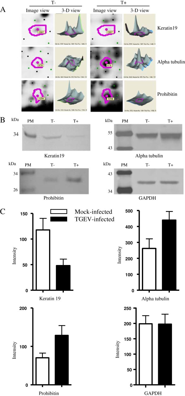
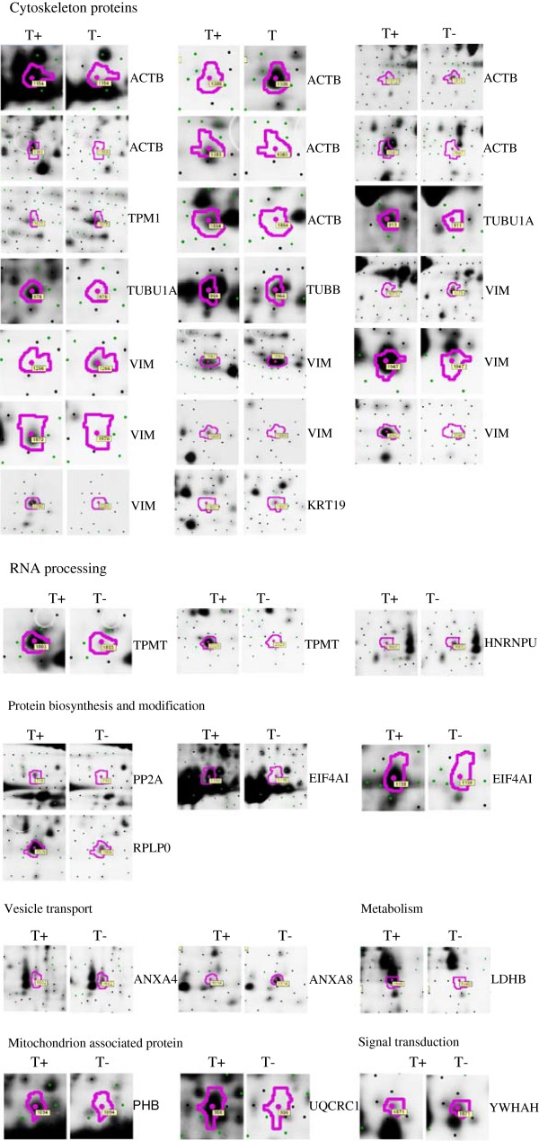
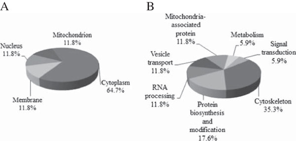
33 differentially expressed protein spots, of which 23 were up-regulated and 10 were down-regulated were identified. All the protein spots were successfully identified. The identified proteins were involved in the regulation of essential processes such as cellular structure and integrity, RNA processing, protein biosynthesis and modification, vesicle transport, signal transduction, and the mitochondrial pathway. Western blot analysis was used to validate the changes of alpha tubulin, keratin 19, and prohibitin during TGEV infection.

CONCLUSIONS

To our knowledge, we have performed the first analysis of the proteomic changes in host cell during TGEV infection. 17 altered cellular proteins that differentially expressed in TGEV infection were identified. The present study provides protein-related information that should be useful for understanding the host cell response to TGEV infection and the underlying mechanism of TGEV replication and pathogenicity.

摘要

背景

传染性胃肠炎冠状病毒(TGEV)是一种引起猪腹泻的肠致病性冠状病毒,与仔猪的高发病率和死亡率有关。关于宿主细胞对 TGEV 感染的比较蛋白质表达的信息仍然有限。在这项研究中,使用二维差异凝胶电泳(2D DIGE)结合 MALDI-TOF-TOF/MS 鉴定的蛋白质组学方法分析了猪睾丸(ST)细胞中 TGEV 感染的细胞蛋白质反应。

结果

鉴定了 33 个差异表达的蛋白质斑点,其中 23 个上调,10 个下调。所有的蛋白斑点都被成功鉴定。鉴定的蛋白质参与了细胞结构和完整性、RNA 处理、蛋白质生物合成和修饰、囊泡运输、信号转导和线粒体途径等重要过程的调节。Western blot 分析用于验证 TGEV 感染过程中 alpha 微管蛋白、角蛋白 19 和抑制素的变化。

结论

据我们所知,我们首次分析了宿主细胞在 TGEV 感染过程中的蛋白质组变化。鉴定了 17 个在 TGEV 感染中差异表达的改变的细胞蛋白。本研究提供了与蛋白质相关的信息,这对于理解宿主细胞对 TGEV 感染的反应以及 TGEV 复制和致病性的潜在机制应该是有用的。

https://cdn.ncbi.nlm.nih.gov/pmc/blobs/39bf/3734006/df0b91ae19e8/1477-5956-11-31-6.jpg
https://cdn.ncbi.nlm.nih.gov/pmc/blobs/39bf/3734006/2517d053f625/1477-5956-11-31-1.jpg
https://cdn.ncbi.nlm.nih.gov/pmc/blobs/39bf/3734006/91535170c837/1477-5956-11-31-2.jpg
https://cdn.ncbi.nlm.nih.gov/pmc/blobs/39bf/3734006/00ac51cddd3f/1477-5956-11-31-3.jpg
https://cdn.ncbi.nlm.nih.gov/pmc/blobs/39bf/3734006/aca13c0557a6/1477-5956-11-31-4.jpg
https://cdn.ncbi.nlm.nih.gov/pmc/blobs/39bf/3734006/76a9d3e6e034/1477-5956-11-31-5.jpg
https://cdn.ncbi.nlm.nih.gov/pmc/blobs/39bf/3734006/df0b91ae19e8/1477-5956-11-31-6.jpg

相似文献

1
Identification of cellular proteome using two-dimensional difference gel electrophoresis in ST cells infected with transmissible gastroenteritis coronavirus.应用二维差异凝胶电泳技术鉴定传染性胃肠炎冠状病毒感染 ST 细胞后的细胞蛋白质组。
Proteome Sci. 2013 Jul 16;11(1):31. doi: 10.1186/1477-5956-11-31.
2
EF1A interacting with nucleocapsid protein of transmissible gastroenteritis coronavirus and plays a role in virus replication.EF1A 与传染性胃肠炎冠状病毒的核衣壳蛋白相互作用,并在病毒复制中发挥作用。
Vet Microbiol. 2014 Aug 27;172(3-4):443-8. doi: 10.1016/j.vetmic.2014.05.034. Epub 2014 Jun 9.
3
Proteome profile of swine testicular cells infected with porcine transmissible gastroenteritis coronavirus.感染猪传染性胃肠炎冠状病毒的猪睾丸细胞的蛋白质组图谱
PLoS One. 2014 Oct 21;9(10):e110647. doi: 10.1371/journal.pone.0110647. eCollection 2014.
4
EIF4A2 interacts with the membrane protein of transmissible gastroenteritis coronavirus and plays a role in virus replication.真核翻译起始因子4A2(EIF4A2)与传染性胃肠炎冠状病毒的膜蛋白相互作用,并在病毒复制中发挥作用。
Res Vet Sci. 2019 Apr;123:39-46. doi: 10.1016/j.rvsc.2018.12.005. Epub 2018 Dec 17.
5
The PERK Arm of the Unfolded Protein Response Negatively Regulates Transmissible Gastroenteritis Virus Replication by Suppressing Protein Translation and Promoting Type I Interferon Production.未折叠蛋白反应的 PERK 臂通过抑制蛋白翻译和促进 I 型干扰素产生来负调控传染性胃肠炎病毒复制。
J Virol. 2018 Jul 17;92(15). doi: 10.1128/JVI.00431-18. Print 2018 Aug 1.
6
Quantitative proteomic analysis reveals that transmissible gastroenteritis virus activates the JAK-STAT1 signaling pathway.定量蛋白质组学分析表明,传染性胃肠炎病毒激活JAK-STAT1信号通路。
J Proteome Res. 2014 Dec 5;13(12):5376-90. doi: 10.1021/pr500173p. Epub 2014 Nov 10.
7
Persistent Transmissible Gastroenteritis Virus Infection Enhances Enterotoxigenic Escherichia coli K88 Adhesion by Promoting Epithelial-Mesenchymal Transition in Intestinal Epithelial Cells.持续性传染性胃肠炎病毒感染通过促进肠上皮细胞的上皮-间质转化增强产肠毒素大肠杆菌K88的黏附。
J Virol. 2017 Oct 13;91(21). doi: 10.1128/JVI.01256-17. Print 2017 Nov 1.
8
Porcine transmissible gastroenteritis virus inhibits NF-κB activity via nonstructural protein 3 to evade host immune system.猪传染性胃肠炎病毒通过非结构蛋白 3 抑制 NF-κB 活性,从而逃避宿主免疫系统。
Virol J. 2019 Aug 5;16(1):97. doi: 10.1186/s12985-019-1206-9.
9
Porcine transmissible gastroenteritis virus nonstructural protein 2 contributes to inflammation via NF-κB activation.猪传染性胃肠炎病毒非结构蛋白 2 通过 NF-κB 激活促进炎症反应。
Virulence. 2018;9(1):1685-1698. doi: 10.1080/21505594.2018.1536632.
10
Interferon gamma inhibits transmissible gastroenteritis virus infection mediated by an IRF1 signaling pathway.干扰素γ通过 IRF1 信号通路抑制传染性胃肠炎病毒感染。
Arch Virol. 2019 Nov;164(11):2659-2669. doi: 10.1007/s00705-019-04362-2. Epub 2019 Aug 5.

引用本文的文献

1
Coronavirus and the Cytoskeleton of Virus-Infected Cells.冠状病毒与病毒感染细胞的细胞骨架
Subcell Biochem. 2023;106:333-364. doi: 10.1007/978-3-031-40086-5_12.
2
The Effects of Swine Coronaviruses on ER Stress, Autophagy, Apoptosis, and Alterations in Cell Morphology.猪冠状病毒对内质网应激、自噬、凋亡及细胞形态改变的影响
Pathogens. 2022 Aug 19;11(8):940. doi: 10.3390/pathogens11080940.
3
Genomewide CRISPR knockout screen identified PLAC8 as an essential factor for SADS-CoVs infection.全基因组 CRISPR 敲除筛选发现 PLAC8 是 SARS-CoV-2 等相关冠状病毒感染的必需因素。

本文引用的文献

1
Identification of porcine serum proteins modified in response to HP-PRRSV HuN4 infection by two-dimensional differential gel electrophoresis.采用二维差异凝胶电泳技术鉴定感染 HP-PRRSV HuN4 后猪血清中发生变化的蛋白。
Vet Microbiol. 2012 Aug 17;158(3-4):237-46. doi: 10.1016/j.vetmic.2012.01.021. Epub 2012 Jan 25.
2
Proteomics analysis of differentially expressed proteins in chicken trachea and kidney after infection with the highly virulent and attenuated coronavirus infectious bronchitis virus in vivo.鸡气管和肾脏感染高致病性和弱毒传染性支气管炎病毒后差异表达蛋白的蛋白质组学分析。
Proteome Sci. 2012 Mar 31;10:24. doi: 10.1186/1477-5956-10-24.
3
Proc Natl Acad Sci U S A. 2022 May 3;119(18):e2118126119. doi: 10.1073/pnas.2118126119. Epub 2022 Apr 27.
4
TMT labeled comparative proteomic analysis reveals spleen active immune responses during type C infected piglet diarrhea.TMT 标记比较蛋白质组学分析揭示了 C 型感染仔猪腹泻期间脾脏的主动免疫反应。
PeerJ. 2022 Apr 4;10:e13006. doi: 10.7717/peerj.13006. eCollection 2022.
5
Cytoskeleton-a crucial key in host cell for coronavirus infection.细胞骨架——冠状病毒感染宿主细胞的关键钥匙。
J Mol Cell Biol. 2020 Jul 1;12(12):968-979. doi: 10.1093/jmcb/mjaa042.
6
Comparative Proteome Analysis of Porcine Jejunum Tissues in Response to a Virulent Strain of Porcine Epidemic Diarrhea Virus and Its Attenuated Strain.猪流行性腹泻病毒强毒株及其弱毒株感染后猪空肠组织的蛋白质组比较分析
Viruses. 2016 Nov 29;8(12):323. doi: 10.3390/v8120323.
7
Tubulins interact with porcine and human S proteins of the genus Alphacoronavirus and support successful assembly and release of infectious viral particles.微管蛋白与甲型冠状病毒属的猪和人S蛋白相互作用,并支持传染性病毒颗粒的成功组装和释放。
Virology. 2016 Oct;497:185-197. doi: 10.1016/j.virol.2016.07.022. Epub 2016 Jul 30.
8
Proteome analysis of porcine epidemic diarrhea virus (PEDV)-infected Vero cells.猪流行性腹泻病毒(PEDV)感染的非洲绿猴肾细胞(Vero细胞)的蛋白质组分析
Proteomics. 2015 Jun;15(11):1819-28. doi: 10.1002/pmic.201400458. Epub 2015 Mar 9.
9
Identification of the interaction between vimentin and nucleocapsid protein of transmissible gastroenteritis virus.鉴定波形蛋白与传染性胃肠炎病毒核衣壳蛋白之间的相互作用。
Virus Res. 2015 Mar 16;200:56-63. doi: 10.1016/j.virusres.2014.12.013. Epub 2014 Dec 20.
10
Looking for a needle in a haystack: Cellular proteins that may interact with the tyrosine-based sorting signal of the TGEV S protein.大海捞针:可能与猪传染性胃肠炎病毒S蛋白基于酪氨酸的分选信号相互作用的细胞蛋白。
Virus Res. 2015 Apr 16;202:3-11. doi: 10.1016/j.virusres.2014.11.029. Epub 2014 Dec 4.
Changes in the nasopharyngeal carcinoma nuclear proteome induced by the EBNA1 protein of Epstein-Barr virus reveal potential roles for EBNA1 in metastasis and oxidative stress responses.
EBV 病毒 EBNA1 蛋白诱导的鼻咽癌核蛋白组改变揭示了 EBNA1 在转移和氧化应激反应中的潜在作用。
J Virol. 2012 Jan;86(1):382-94. doi: 10.1128/JVI.05648-11. Epub 2011 Oct 19.
4
Proteomic analysis of an Aedes albopictus cell line infected with Dengue serotypes 1 and 3 viruses.登革病毒 1 型和 3 型感染白纹伊蚊细胞系的蛋白质组学分析。
Parasit Vectors. 2011 Jul 18;4:138. doi: 10.1186/1756-3305-4-138.
5
Proteomic analysis of chicken embryonic trachea and kidney tissues after infection in ovo by avian infectious bronchitis coronavirus.鸡胚感染禽传染性支气管炎冠状病毒后气管和肾脏组织的蛋白质组学分析。
Proteome Sci. 2011 Mar 8;9:11. doi: 10.1186/1477-5956-9-11.
6
Identification of host factors involved in coronavirus replication by quantitative proteomics analysis.通过定量蛋白质组学分析鉴定冠状病毒复制过程中的宿主因子。
Proteomics. 2011 Jan;11(1):64-80. doi: 10.1002/pmic.201000309. Epub 2010 Dec 6.
7
HCV inhibits antigen processing and presentation and induces oxidative stress response in gastric mucosa.丙型肝炎病毒抑制抗原加工和呈递,并诱导胃黏膜氧化应激反应。
Proteomics Clin Appl. 2008 Sep;2(9):1290-9. doi: 10.1002/prca.200800059. Epub 2008 Jul 21.
8
Papain-like protease 1 from transmissible gastroenteritis virus: crystal structure and enzymatic activity toward viral and cellular substrates.传染性胃肠炎病毒的木瓜蛋白酶样蛋白酶 1:晶体结构及对病毒和细胞底物的酶活性。
J Virol. 2010 Oct;84(19):10063-73. doi: 10.1128/JVI.00898-10. Epub 2010 Jul 28.
9
Quantitative proteomics using stable isotope labeling with amino acids in cell culture reveals changes in the cytoplasmic, nuclear, and nucleolar proteomes in Vero cells infected with the coronavirus infectious bronchitis virus.稳定同位素标记与细胞培养氨基酸标记的定量蛋白质组学揭示了冠状病毒传染性支气管炎病毒感染的vero 细胞中细胞质、核和核仁蛋白质组的变化。
Mol Cell Proteomics. 2010 Sep;9(9):1920-36. doi: 10.1074/mcp.M900345-MCP200. Epub 2010 May 13.
10
Molecular characterization of a Chinese vaccine strain of transmissible gastroenteritis virus: mutations that may contribute to attenuation.中国猪传染性胃肠炎病毒疫苗株的分子特征:可能导致病毒减毒的突变
Virus Genes. 2010 Jun;40(3):403-9. doi: 10.1007/s11262-010-0467-6. Epub 2010 Mar 13.