• 文献检索
  • 文档翻译
  • 深度研究
  • 学术资讯
  • Suppr Zotero 插件Zotero 插件
  • 邀请有礼
  • 套餐&价格
  • 历史记录
应用&插件
Suppr Zotero 插件Zotero 插件浏览器插件Mac 客户端Windows 客户端微信小程序
定价
高级版会员购买积分包购买API积分包
服务
文献检索文档翻译深度研究API 文档MCP 服务
关于我们
关于 Suppr公司介绍联系我们用户协议隐私条款
关注我们

Suppr 超能文献

核心技术专利:CN118964589B侵权必究
粤ICP备2023148730 号-1Suppr @ 2026

文献检索

告别复杂PubMed语法,用中文像聊天一样搜索,搜遍4000万医学文献。AI智能推荐,让科研检索更轻松。

立即免费搜索

文件翻译

保留排版,准确专业,支持PDF/Word/PPT等文件格式,支持 12+语言互译。

免费翻译文档

深度研究

AI帮你快速写综述,25分钟生成高质量综述,智能提取关键信息,辅助科研写作。

立即免费体验

新生小鼠皮层轴突和树突结构的体内成像。

In vivo imaging of axonal and dendritic structures in neonatal mouse cortex.

作者信息

Cruz-Martin Alberto, Portera-Cailliau Carlos

出版信息

Cold Spring Harb Protoc. 2014 Jan 1;2014(1):57-64. doi: 10.1101/pdb.prot080150.

DOI:10.1101/pdb.prot080150
PMID:24371322
原文链接:https://pmc.ncbi.nlm.nih.gov/articles/PMC9809208/
Abstract

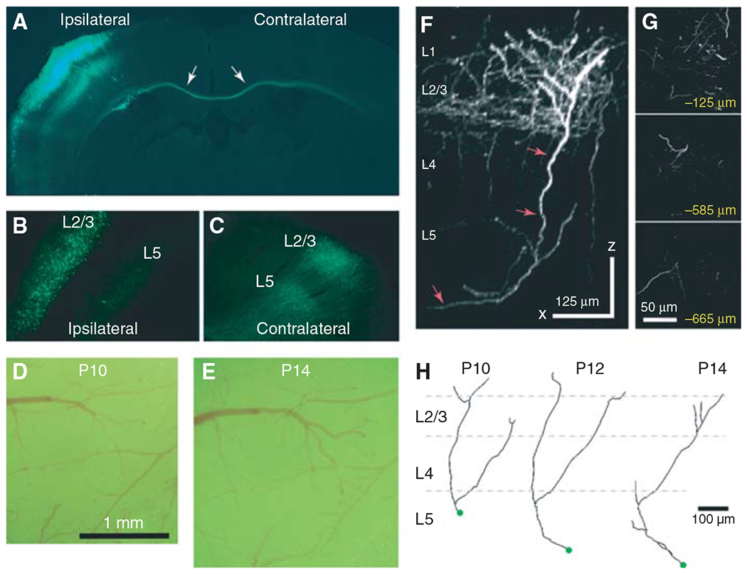
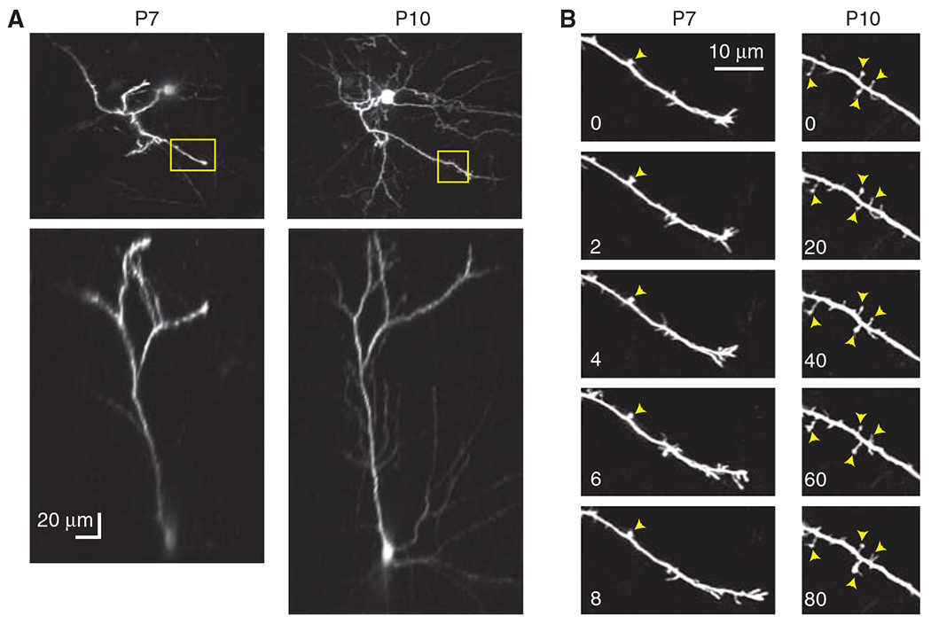
Time-lapse in vivo imaging of neuronal structures is critical for understanding the assembly of neural circuits during development. Imaging developing neurons in vivo can be performed with relative ease in lower vertebrates, but ideally, one would also like to image the developing mammalian brain. In vivo chronic imaging of mice is particularly desirable because of the availability of transgenic lines that model human neuropsychiatric disease or those that allow cell- or region-specific expression of fluorescent proteins (e.g., green fluorescent protein [GFP], channelrhodopsins, and genetically encoded calcium indicators). Unfortunately, although chronic imaging of neural structures in adult mice that express GFP is now commonplace, similar approaches in neonatal mice face several additional challenges. First, the small size of the animal complicates the cranial window surgery. Second, there is a tendency for dams to cannibalize pups with head caps. Third, the head cap can impede the normal growth of the skull in neonates, which can limit the duration of imaging. Here, we describe a method for implanting chronic glass-covered cranial windows in the skulls of early postnatal mice through which axonal and dendritic structures can be imaged in vivo over a period of hours or even days.

摘要

对神经元结构进行延时活体成像对于理解发育过程中神经回路的组装至关重要。在低等脊椎动物中,对发育中的神经元进行活体成像相对容易,但理想情况下,人们也希望对发育中的哺乳动物大脑进行成像。由于存在模拟人类神经精神疾病的转基因品系,或允许荧光蛋白(如绿色荧光蛋白[GFP]、通道视紫红质和基因编码钙指示剂)进行细胞或区域特异性表达的转基因品系,对小鼠进行活体长期成像尤为可取。不幸的是,尽管目前对表达GFP的成年小鼠的神经结构进行长期成像已很常见,但在新生小鼠中采用类似方法面临一些额外挑战。首先,动物体型小使颅骨开窗手术变得复杂。其次,母鼠有吃掉带帽幼崽的倾向。第三,头帽会阻碍新生小鼠颅骨的正常生长,这可能会限制成像持续时间。在此,我们描述一种在出生后早期小鼠颅骨中植入慢性玻璃覆盖颅骨窗的方法,通过该窗口可在数小时甚至数天内对轴突和树突结构进行活体成像。

https://cdn.ncbi.nlm.nih.gov/pmc/blobs/5bfe/9809208/b2f96460ab4c/nihms-1857462-f0002.jpg
https://cdn.ncbi.nlm.nih.gov/pmc/blobs/5bfe/9809208/688aa3917eb6/nihms-1857462-f0001.jpg
https://cdn.ncbi.nlm.nih.gov/pmc/blobs/5bfe/9809208/b2f96460ab4c/nihms-1857462-f0002.jpg
https://cdn.ncbi.nlm.nih.gov/pmc/blobs/5bfe/9809208/688aa3917eb6/nihms-1857462-f0001.jpg
https://cdn.ncbi.nlm.nih.gov/pmc/blobs/5bfe/9809208/b2f96460ab4c/nihms-1857462-f0002.jpg

相似文献

1
In vivo imaging of axonal and dendritic structures in neonatal mouse cortex.新生小鼠皮层轴突和树突结构的体内成像。
Cold Spring Harb Protoc. 2014 Jan 1;2014(1):57-64. doi: 10.1101/pdb.prot080150.
2
NMDAR-regulated dynamics of layer 4 neuronal dendrites during thalamocortical reorganization in neonates.NMDA 受体调控的新生儿丘脑皮质重组过程中层 4 神经元树突的动态变化。
Neuron. 2014 Apr 16;82(2):365-79. doi: 10.1016/j.neuron.2014.02.026. Epub 2014 Mar 27.
3
Depolarization stimulates lamellipodia formation and axonal but not dendritic branching in cultured rat cerebral cortex neurons.去极化刺激培养的大鼠大脑皮层神经元中片状伪足的形成和轴突分支,但不刺激树突分支。
Brain Res Dev Brain Res. 1998 Jun 15;108(1-2):205-16. doi: 10.1016/s0165-3806(98)00050-9.
4
Skin suturing and cortical surface viral infusion improves imaging of neuronal ensemble activity with head-mounted miniature microscopes.皮肤缝合和皮质表面病毒注射有助于使用头戴式微型显微镜对神经元集合活动进行成像。
J Neurosci Methods. 2017 Nov 1;291:238-248. doi: 10.1016/j.jneumeth.2017.08.016. Epub 2017 Aug 19.
5
Dendrites In Vitro and In Vivo Contain Microtubules of Opposite Polarity and Axon Formation Correlates with Uniform Plus-End-Out Microtubule Orientation.体外和体内的树突含有极性相反的微管,轴突形成与统一的正端向外微管方向相关。
J Neurosci. 2016 Jan 27;36(4):1071-85. doi: 10.1523/JNEUROSCI.2430-15.2016.
6
Nonuniform alteration of dendritic development in the cerebral cortex following prenatal cocaine exposure.产前接触可卡因后大脑皮质树突发育的不均匀改变。
Cereb Cortex. 1996 May-Jun;6(3):431-45. doi: 10.1093/cercor/6.3.431.
7
Time-Lapse Imaging of Neuronal Arborization using Sparse Adeno-Associated Virus Labeling of Genetically Targeted Retinal Cell Populations.使用基因靶向视网膜细胞群体的稀疏腺相关病毒标记进行神经元树突分支的延时成像。
J Vis Exp. 2021 Mar 19(169). doi: 10.3791/62308.
8
In vivo postnatal electroporation and time-lapse imaging of neuroblast migration in mouse acute brain slices.小鼠急性脑片中神经母细胞迁移的体内产后电穿孔及延时成像
J Vis Exp. 2013 Nov 25(81):50905. doi: 10.3791/50905.
9
In vivo two-photon imaging of experience-dependent molecular changes in cortical neurons.皮层神经元中经验依赖性分子变化的体内双光子成像
J Vis Exp. 2013 Jan 5(71):50148. doi: 10.3791/50148.
10
Laser nanosurgery of cerebellar axons in vivo.体内小脑轴突的激光纳米手术
J Vis Exp. 2014 Jul 28(89):e51371. doi: 10.3791/51371.

引用本文的文献

1
A pipeline for STED super-resolution imaging and Imaris analysis of nanoscale synapse organization in mouse cortical brain slices.用于 STED 超分辨率成像和 Imaris 分析小鼠皮质脑片纳米级突触组织的流水线。
STAR Protoc. 2023 Dec 15;4(4):102707. doi: 10.1016/j.xpro.2023.102707. Epub 2023 Nov 9.
2
The Shape of μ-How Morphological Analyses Shape the Study of Microglia.μ-如何:形态学分析如何塑造小胶质细胞研究的形态
Front Cell Neurosci. 2022 Jun 29;16:942462. doi: 10.3389/fncel.2022.942462. eCollection 2022.
3
Assessing Microglial Dynamics by Live Imaging.

本文引用的文献

1
Delayed stabilization of dendritic spines in fragile X mice.脆性 X 小鼠树突棘的延迟稳定。
J Neurosci. 2010 Jun 9;30(23):7793-803. doi: 10.1523/JNEUROSCI.0577-10.2010.
2
Internally mediated developmental desynchronization of neocortical network activity.新皮质网络活动的内部介导发育不同步
J Neurosci. 2009 Sep 2;29(35):10890-9. doi: 10.1523/JNEUROSCI.2012-09.2009.
3
Long-term, high-resolution imaging in the mouse neocortex through a chronic cranial window.通过慢性颅骨视窗对小鼠新皮层进行长期、高分辨率成像。
通过实时成像评估小胶质细胞动力学。
Front Immunol. 2021 Mar 8;12:617564. doi: 10.3389/fimmu.2021.617564. eCollection 2021.
4
Through the looking glass: A review of cranial window technology for optical access to the brain.透过镜子:颅窗技术用于大脑光学接入的回顾。
J Neurosci Methods. 2021 Apr 15;354:109100. doi: 10.1016/j.jneumeth.2021.109100. Epub 2021 Feb 15.
5
Spatio-temporal dynamics of neocortical presynaptic terminal development using multi-photon imaging of the corpus callosum in vivo.利用体内大脑胼胝体的多光子成像技术研究新皮层突触前末梢发育的时空动态。
Sci Rep. 2019 Oct 1;9(1):14028. doi: 10.1038/s41598-019-50431-6.
6
Skull optical clearing window for imaging of the mouse cortex at synaptic resolution.用于在突触分辨率下对小鼠皮层进行成像的颅骨光学透明窗口。
Light Sci Appl. 2018 Feb 23;7:17153. doi: 10.1038/lsa.2017.153. eCollection 2018.
7
Dendritic Spines in Early Postnatal Fragile X Mice Are Insensitive to Novel Sensory Experience.早期产后脆性 X 小鼠的树突棘对新感觉体验不敏感。
J Neurosci. 2019 Jan 16;39(3):412-419. doi: 10.1523/JNEUROSCI.1734-18.2018. Epub 2018 Dec 6.
8
Design and implementation of multi-signal and time-varying neural reconstructions.多信号和时变神经重构的设计与实现。
Sci Data. 2018 Jan 23;5:170207. doi: 10.1038/sdata.2017.207.
9
Comparison of intravital thinned skull and cranial window approaches to study CNS immunobiology in the mouse cortex.用于研究小鼠皮质中枢神经系统免疫生物学的活体减薄颅骨法与颅骨开窗法的比较。
Intravital. 2014 May;3(2). doi: 10.4161/intv.29728.
10
Two-photon excitation microscopy and its applications in neuroscience.双光子激发显微镜及其在神经科学中的应用。
Methods Mol Biol. 2015;1251:25-42. doi: 10.1007/978-1-4939-2080-8_2.
Nat Protoc. 2009;4(8):1128-44. doi: 10.1038/nprot.2009.89. Epub 2009 Jul 16.
4
A method for 2-photon imaging of blood flow in the neocortex through a cranial window.一种通过颅骨视窗对新皮层血流进行双光子成像的方法。
J Vis Exp. 2008 Feb 25(12):678. doi: 10.3791/678.
5
A craniotomy surgery procedure for chronic brain imaging.一种用于慢性脑成像的开颅手术程序。
J Vis Exp. 2008 Feb 15(12):680. doi: 10.3791/680.
6
Rapid redistribution of synaptic PSD-95 in the neocortex in vivo.体内新皮质中突触后密度蛋白95(PSD-95)的快速重新分布。
PLoS Biol. 2006 Nov;4(11):e370. doi: 10.1371/journal.pbio.0040370.
7
Diverse modes of axon elaboration in the developing neocortex.发育中的新皮层中轴突形成的多种模式。
PLoS Biol. 2005 Aug;3(8):e272. doi: 10.1371/journal.pbio.0030272. Epub 2005 Jul 26.
8
Electroporation and RNA interference in the rodent retina in vivo and in vitro.体内和体外啮齿动物视网膜中的电穿孔和RNA干扰
Proc Natl Acad Sci U S A. 2004 Jan 6;101(1):16-22. doi: 10.1073/pnas.2235688100. Epub 2003 Nov 5.
9
ScanImage: flexible software for operating laser scanning microscopes.ScanImage:用于操作激光扫描显微镜的灵活软件。
Biomed Eng Online. 2003 May 17;2:13. doi: 10.1186/1475-925X-2-13.
10
Efficient gene transfer into the embryonic mouse brain using in vivo electroporation.利用体内电穿孔技术将基因高效导入胚胎小鼠大脑。
Dev Biol. 2001 Dec 1;240(1):237-46. doi: 10.1006/dbio.2001.0439.