Morganti Josh M, Jopson Timothy D, Liu Sharon, Gupta Nalin, Rosi Susanna
Brain and Spinal Injury Center, University of California San Francisco, San Francisco, California, United States of America; Departments of Physical Therapy and Rehabilitation Science, University of California San Francisco, San Francisco, California, United States of America.
Neurological Surgery, University of California San Francisco, San Francisco, California, United States of America.
PLoS One. 2014 Apr 2;9(4):e93650. doi: 10.1371/journal.pone.0093650. eCollection 2014.
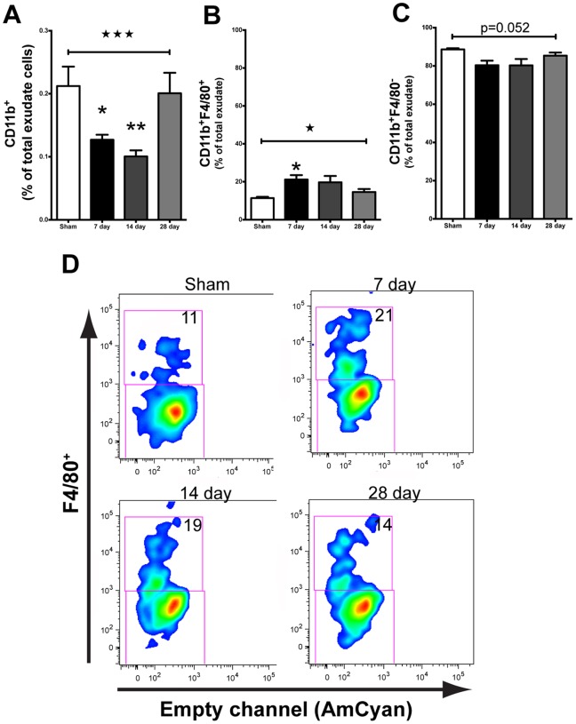
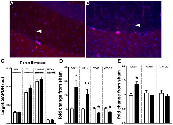
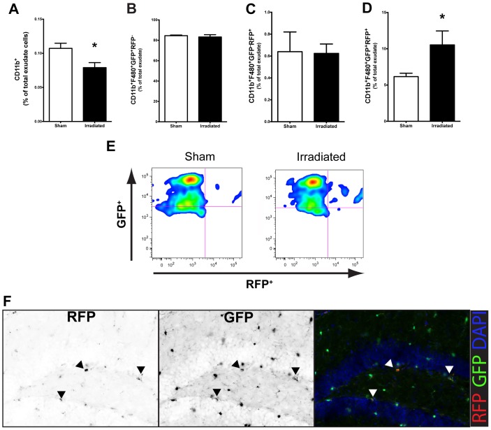
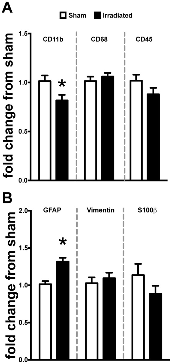
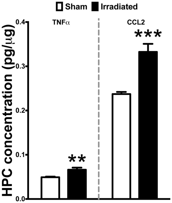
Therapeutic irradiation is commonly used to treat primary or metastatic central nervous system tumors. It is believed that activation of neuroinflammatory signaling pathways contributes to the development of common adverse effects, which may ultimately contribute to cognitive dysfunction. Recent studies identified the chemokine (C-C motif) receptor (CCR2), constitutively expressed by cells of the monocyte-macrophage lineage, as a mediator of cognitive impairments induced by irradiation. In the present study we utilized a unique reporter mouse (CCR2(RFP/+)CX3CR1(GFP/+)) to accurately delineate the resident (CX3CR1+) versus peripheral (CCR2+) innate immune response in the brain following cranial irradiation. Our results demonstrate that a single dose of 10Gy cranial γ-irradiation induced a significant decrease in the percentage of resident microglia, while inducing an increase in the infiltration of peripherally derived CCR2+ macrophages. Although reduced in percentage, there was a significant increase in F4/80+ activated macrophages in irradiated animals compared to sham. Moreover, we found that there were altered levels of pro-inflammatory cytokines, chemokines, adhesion molecules, and growth factors in the hippocampi of wild type irradiated mice as compared to sham. All of these molecules are implicated in the recruitment, adhesion, and migration of peripheral monocytes to injured tissue. Importantly, there were no measureable changes in the expression of multiple markers associated with blood-brain barrier integrity; implicating the infiltration of peripheral CCR2+ macrophages may be due to inflammatory induced chemotactic signaling. Cumulatively, these data provide evidence that therapeutic levels of cranial radiation are sufficient to alter the brain's homeostatic balance and permit the influx of peripherally-derived CCR2+ macrophages as well as the regional susceptibility of the hippocampal formation to ionizing radiation.
治疗性放射常用于治疗原发性或转移性中枢神经系统肿瘤。据信,神经炎症信号通路的激活会导致常见不良反应的发生,最终可能导致认知功能障碍。最近的研究发现,趋化因子(C-C基序)受体(CCR2)由单核巨噬细胞谱系细胞组成性表达,是辐射诱导认知障碍的介质。在本研究中,我们利用一种独特的报告小鼠(CCR2(RFP/+)CX3CR1(GFP/+))来准确描绘颅脑照射后大脑中驻留(CX3CR1+)与外周(CCR2+)固有免疫反应。我们的结果表明,单次10Gy颅脑γ射线照射导致驻留小胶质细胞百分比显著下降,同时外周来源的CCR2+巨噬细胞浸润增加。与假手术组相比,照射动物中F4/80+活化巨噬细胞的百分比虽然降低,但数量显著增加。此外,我们发现与假手术组相比,野生型照射小鼠海马中促炎细胞因子、趋化因子、黏附分子和生长因子的水平发生了改变。所有这些分子都与外周单核细胞向损伤组织的募集、黏附和迁移有关。重要的是,与血脑屏障完整性相关的多个标志物的表达没有可测量的变化;这表明外周CCR2+巨噬细胞的浸润可能是由于炎症诱导的趋化信号。累积起来,这些数据提供了证据,表明颅脑放疗的治疗水平足以改变大脑的稳态平衡,允许外周来源的CCR2+巨噬细胞流入,以及海马结构对电离辐射的区域易感性。