Reddy Vandana, Bhagwath Sundeep S, Reddy Munish
Department of Oral Pathology and Microbiology, Subharti Dental College, Meerut, Uttar Pradesh, India.
Department of Orthodontics, Subharti Dental College, Meerut, Uttar Pradesh, India.
Dent Res J (Isfahan). 2014 Mar;11(2):187-92.
The aim of this study was to quantify the number of mast cells in focal reactive hyperplastic lesions of the oral cavity and to compare these two number of mast cells in normal gingival tissues and to correlate their presence with the state of connective tissue changes in reactive lesions and probably suggest a role for mast cells in these lesions.
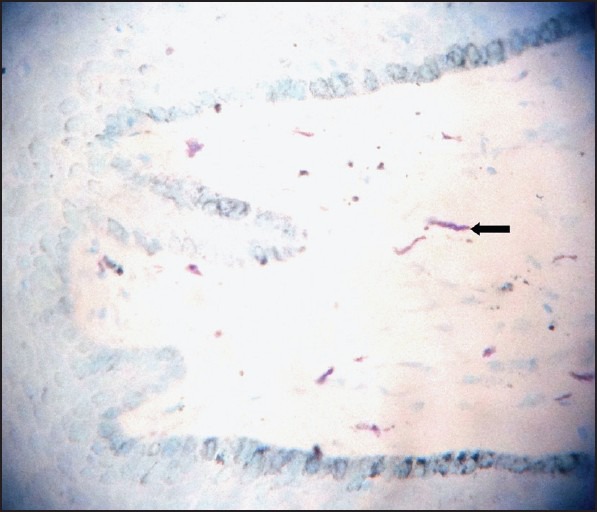
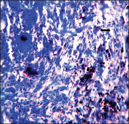
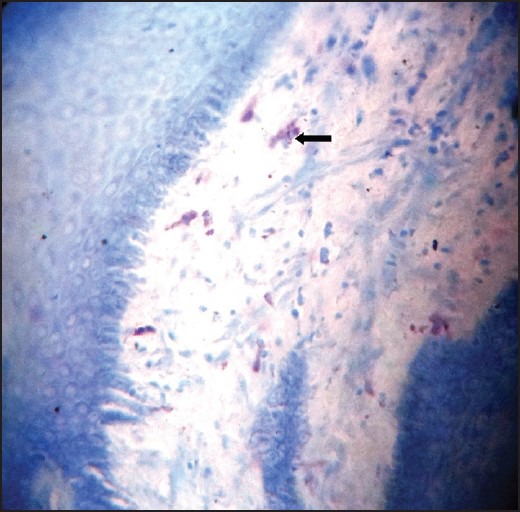
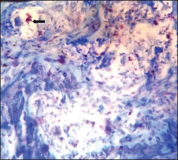
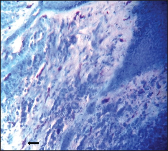
Patient records were retrieved during a 10 year period from 2001 to 2010. Data of all reactive hyperplasias namely focal fibrous hyperplasia, pyogenic granuloma (PG), peripheral ossifying fibroma (POF) and peripheral giant cell granuloma (PGCG) were reviewed and 10 cases seen in the gingiva were selected for each category and stained with 1% toluidine blue for mast cells. Statistical analysis was applied to see the significant differences between the groups and with the normal gingival tissue. One-way ANOVA-F and unpaired t-test was applied and significant differences were seen between the groups at 5% level of significance.
In this study, mast cell count was maximum in POF and fibrous hyperplasia (FH) followed by cases of PG and PGCG.
The number of mast cells was more numerous in POF and FH suggesting that mast cell activation is a characteristic feature of chronic inflammation, a condition that may lead to fibrosis as a result of increased collagen synthesis by fibroblasts.
本研究的目的是量化口腔局灶性反应性增生性病变中肥大细胞的数量,比较其与正常牙龈组织中肥大细胞数量的差异,并将它们的存在与反应性病变中结缔组织变化的状态相关联,可能提示肥大细胞在这些病变中的作用。
检索2001年至2010年10年间的患者记录。回顾所有反应性增生的数据,即局灶性纤维增生、化脓性肉芽肿(PG)、外周骨化性纤维瘤(POF)和外周巨细胞肉芽肿(PGCG),并为每个类别选择10例牙龈病变病例,用1%甲苯胺蓝对肥大细胞进行染色。应用统计分析来观察各组之间以及与正常牙龈组织之间的显著差异。采用单因素方差分析-F和非配对t检验,在5%的显著性水平上观察到各组之间存在显著差异。
在本研究中,POF和纤维增生(FH)中的肥大细胞计数最高,其次是PG和PGCG病例。
POF和FH中的肥大细胞数量更多,提示肥大细胞活化是慢性炎症的一个特征,这种情况可能由于成纤维细胞胶原合成增加而导致纤维化。