• 文献检索
  • 文档翻译
  • 深度研究
  • 学术资讯
  • Suppr Zotero 插件Zotero 插件
  • 邀请有礼
  • 套餐&价格
  • 历史记录
应用&插件
Suppr Zotero 插件Zotero 插件浏览器插件Mac 客户端Windows 客户端微信小程序
定价
高级版会员购买积分包购买API积分包
服务
文献检索文档翻译深度研究API 文档MCP 服务
关于我们
关于 Suppr公司介绍联系我们用户协议隐私条款
关注我们

Suppr 超能文献

核心技术专利:CN118964589B侵权必究
粤ICP备2023148730 号-1Suppr @ 2026

文献检索

告别复杂PubMed语法,用中文像聊天一样搜索,搜遍4000万医学文献。AI智能推荐,让科研检索更轻松。

立即免费搜索

文件翻译

保留排版,准确专业,支持PDF/Word/PPT等文件格式,支持 12+语言互译。

免费翻译文档

深度研究

AI帮你快速写综述,25分钟生成高质量综述,智能提取关键信息,辅助科研写作。

立即免费体验

口腔反应性病变中肥大细胞计数及微血管密度的评估

Evaluation of mast cell counts and microvessel density in reactive lesions of the oral cavity.

作者信息

Kouhsoltani Maryam, Moradzadeh Khiavi Monir, Tahamtan Shabnam

机构信息

Dental and Periodontal Research Center and Faculty of Dentistry, Tabriz University of Medical Sciences, Tabriz, Iran.

Department of Oral and Maxillofacial Pathology, Faculty of Dentistry, Tehran University of Medical Sciences, Tehran, Iran.

出版信息

J Dent Res Dent Clin Dent Prospects. 2016 Fall;10(4):241-246. doi: 10.15171/joddd.2016.038. Epub 2016 Dec 21.

DOI:10.15171/joddd.2016.038
PMID:28096950
原文链接:https://pmc.ncbi.nlm.nih.gov/articles/PMC5237671/
Abstract

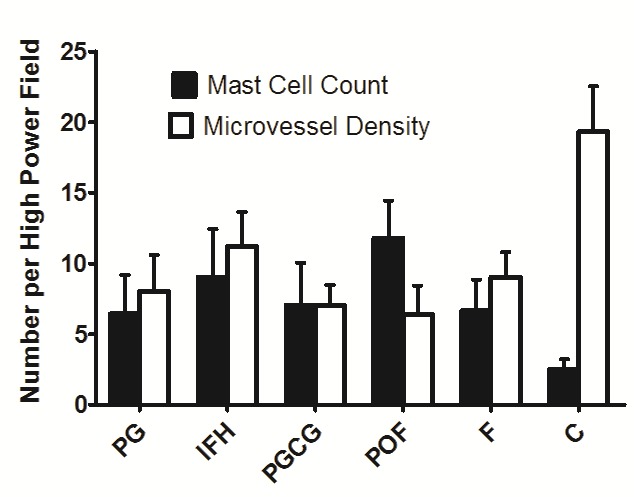
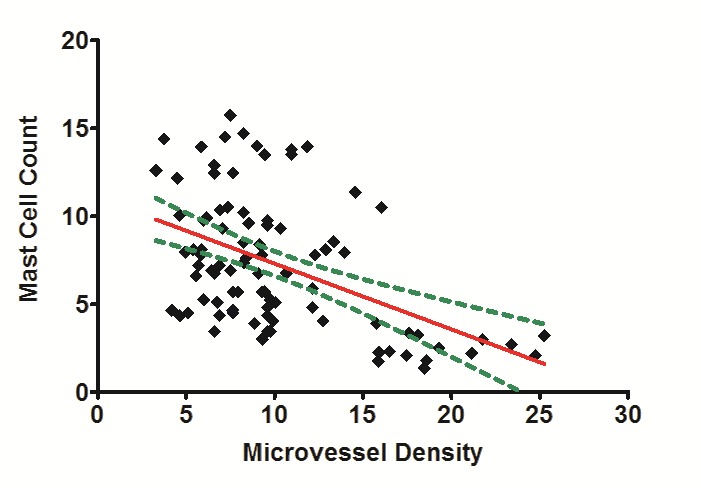
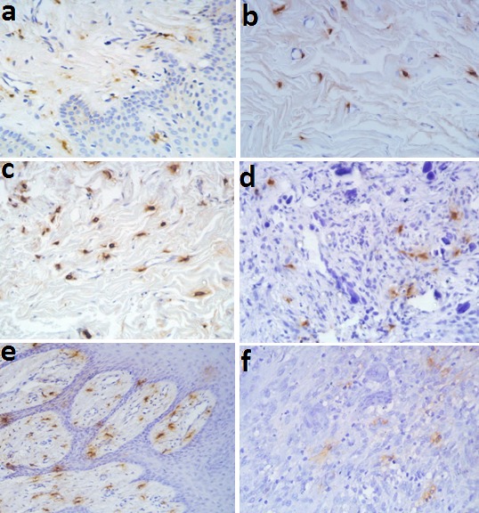
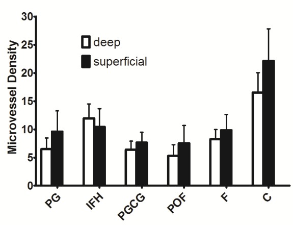
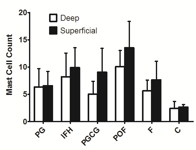
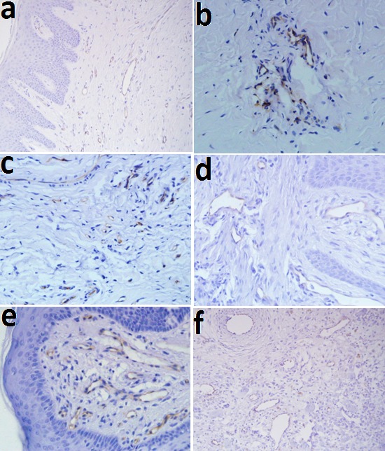
Reliable immunohistochemical assays to assess the definitive role of mast cells (MCs) and angiogenesis in the pathogenesis of oral reactive lesions are generally not available. The aim of the present study was to evaluate mast cell counts (MCC) and microvessel density (MVD) in oral reactive lesions and determine the correlation between MCC and MVD. Seventy-five cases of reactive lesions of the oral cavity, including pyogenic granuloma, fibroma, peripheral giant cell granuloma, inflammatory fibrous hyperplasia, peripheral ossifying fibroma (15 for each category) were immunohisto-chemically stained with MC tryptase and CD31. Fifteen cases of normal gingival tissue were considered as the control group. The mean MCC and MVD in superficial and deep connective tissues were assessed and total MCC and MVD was computed for each lesion. . Statistically significant differences were observed in MCC and MVD between the study groups (P < 0.001). MC tryptase and CD31 expression increased in the superficial connective tissue of each lesion in comparison to the deep con-nective tissue. A significant negative correlation was not found between MCC and MVD in oral reactive lesions (P < 0.001, r = -0.458). Although MCs were present in the reactive lesions of the oral cavity, a direct correlation between MCC and MVD was not found in these lesions. Therefore, a significant interaction between MCs and endothelial cells and an active role for MCs in the growth of oral reactive lesions was not found in this study.

摘要

目前尚无可靠的免疫组织化学检测方法来评估肥大细胞(MCs)和血管生成在口腔反应性病变发病机制中的决定性作用。本研究的目的是评估口腔反应性病变中的肥大细胞计数(MCC)和微血管密度(MVD),并确定MCC与MVD之间的相关性。对75例口腔反应性病变进行免疫组织化学染色,包括化脓性肉芽肿、纤维瘤、外周巨细胞肉芽肿、炎性纤维增生、外周骨化性纤维瘤(各15例),采用MC类胰蛋白酶和CD31进行染色。15例正常牙龈组织作为对照组。评估浅、深结缔组织中的平均MCC和MVD,并计算每个病变的总MCC和MVD。研究组之间的MCC和MVD存在统计学显著差异(P < 0.001)。与深结缔组织相比,每个病变的浅结缔组织中MC类胰蛋白酶和CD31表达增加。口腔反应性病变中MCC与MVD之间未发现显著负相关(P < 0.001,r = -0.458)。虽然MCs存在于口腔反应性病变中,但在这些病变中未发现MCC与MVD之间的直接相关性。因此,本研究未发现MCs与内皮细胞之间存在显著相互作用,也未发现MCs在口腔反应性病变生长中起积极作用。

https://cdn.ncbi.nlm.nih.gov/pmc/blobs/1749/5237671/c8921143f121/joddd-10-241-g006.jpg
https://cdn.ncbi.nlm.nih.gov/pmc/blobs/1749/5237671/5c4d6a785f7a/joddd-10-241-g001.jpg
https://cdn.ncbi.nlm.nih.gov/pmc/blobs/1749/5237671/81fb0587852b/joddd-10-241-g002.jpg
https://cdn.ncbi.nlm.nih.gov/pmc/blobs/1749/5237671/e2c905b66c05/joddd-10-241-g003.jpg
https://cdn.ncbi.nlm.nih.gov/pmc/blobs/1749/5237671/79175a0eb4f8/joddd-10-241-g004.jpg
https://cdn.ncbi.nlm.nih.gov/pmc/blobs/1749/5237671/85d165c877b7/joddd-10-241-g005.jpg
https://cdn.ncbi.nlm.nih.gov/pmc/blobs/1749/5237671/c8921143f121/joddd-10-241-g006.jpg

相似文献

1
Evaluation of mast cell counts and microvessel density in reactive lesions of the oral cavity.口腔反应性病变中肥大细胞计数及微血管密度的评估
J Dent Res Dent Clin Dent Prospects. 2016 Fall;10(4):241-246. doi: 10.15171/joddd.2016.038. Epub 2016 Dec 21.
2
Immunohistochemical Expression of Mast Cell in Oral Reactive Lesions.肥大细胞在口腔反应性病变中的免疫组化表达
J Dent (Shiraz). 2015 Mar;16(1 Suppl):10-4.
3
Involvement of mast cells and microvessels density in reactive lesions of oral cavity: A comparative immunohistochemical study.肥大细胞和微血管密度在口腔反应性病变中的作用:一项比较性免疫组织化学研究。
Pathol Res Pract. 2016 Sep;212(9):761-6. doi: 10.1016/j.prp.2016.05.007. Epub 2016 May 18.
4
Comparative Study of Mast Cell Count in Oral Reactive Lesions and Its Association with Inflammation.口腔反应性病变中肥大细胞计数的比较研究及其与炎症的关联
Turk Patoloji Derg. 2016;32(1):22-6. doi: 10.5146/tjpath.2015.01338.
5
Mast cell count in oral reactive lesions: A histochemical study.口腔反应性病变中的肥大细胞计数:一项组织化学研究。
Dent Res J (Isfahan). 2014 Mar;11(2):187-92.
6
Comparison of mast cells count in oral reactive lesions.口腔反应性病变中肥大细胞计数的比较。
Pathol Res Pract. 2010 Mar 15;206(3):151-5. doi: 10.1016/j.prp.2009.10.006. Epub 2010 Jan 21.
7
Assessment of reactive gingival lesions of oral cavity: A histopathological study.口腔反应性牙龈病变的评估:一项组织病理学研究。
J Oral Maxillofac Pathol. 2017 Jan-Apr;21(1):180. doi: 10.4103/jomfp.JOMFP_23_16.
8
Quantification of mast cells in nonreactive and reactive lesions of gingiva: A comparative study using toluidine blue and immunohistochemical marker mast cell tryptase.牙龈非反应性和反应性病变中肥大细胞的定量分析:一项使用甲苯胺蓝和免疫组化标志物肥大细胞类胰蛋白酶的比较研究。
J Oral Maxillofac Pathol. 2021 Sep-Dec;25(3):550-551. doi: 10.4103/jomfp.JOMFP_267_19. Epub 2022 Jan 11.
9
Mast cells and angiogenesis in malignant and premalignant oral lesions: An immunohistochemical study.口腔恶性及癌前病变中的肥大细胞与血管生成:一项免疫组织化学研究
J Oral Maxillofac Pathol. 2017 May-Aug;21(2):229-238. doi: 10.4103/jomfp.JOMFP_111_15.
10
Detection and quantification of mast cell, vascular endothelial growth factor, and microvessel density in human inflammatory periapical cysts and granulomas.检测和定量分析人炎症性根尖囊肿和肉芽肿中的肥大细胞、血管内皮生长因子和微血管密度。
Int Endod J. 2012 Sep;45(9):859-64. doi: 10.1111/j.1365-2591.2012.02043.x. Epub 2012 Apr 5.

引用本文的文献

1
Oral Pyogenic Granuloma: A Narrative Review.口腔化脓性肉芽肿:一篇叙述性综述。
Int J Mol Sci. 2023 Nov 28;24(23):16885. doi: 10.3390/ijms242316885.

本文引用的文献

1
Mast cell count in oral reactive lesions: A histochemical study.口腔反应性病变中的肥大细胞计数:一项组织化学研究。
Dent Res J (Isfahan). 2014 Mar;11(2):187-92.
2
A lack of correlation between mast cells, angiogenesis, and outcome in non-small cell lung cancer.肥大细胞、血管生成与非小细胞肺癌预后之间缺乏相关性。
Exp Lung Res. 2012 Aug;38(6):281-5. doi: 10.3109/01902148.2012.686559. Epub 2012 May 30.
3
Evaluation of mast cells in periapical cysts, dentigerous cysts, and keratocystic odontogenic tumors.评估根尖周囊肿、含牙囊肿和角化囊肿中的肥大细胞。
J Oral Pathol Med. 2012 Sep;41(8):630-6. doi: 10.1111/j.1600-0714.2012.01126.x. Epub 2012 Jan 27.
4
Microvessel density in follicular cysts, keratocystic odontogenic tumours and ameloblastomas.滤泡囊肿、牙源性角化囊性瘤及成釉细胞瘤中的微血管密度
Asian Pac J Cancer Prev. 2011;12(2):351-6.
5
Immunohistochemical expression of mast cell tryptase in giant cell fibroma and inflammatory fibrous hyperplasia of the oral mucosa.肥大细胞类胰蛋白酶在口腔黏膜巨细胞纤维瘤和炎性纤维性增生中的免疫组织化学表达。
Arch Oral Biol. 2011 Mar;56(3):231-7. doi: 10.1016/j.archoralbio.2010.09.020. Epub 2010 Oct 29.
6
Mast cell density and angiogenesis in oral dysplastic epithelium and low- and high-grade oral squamous cell carcinoma.口腔发育不良上皮和低级别及高级别口腔鳞状细胞癌中的肥大细胞密度和血管生成。
Acta Odontol Scand. 2010 Sep;68(5):300-4. doi: 10.3109/00016357.2010.494622.
7
Comparison of mast cells count in oral reactive lesions.口腔反应性病变中肥大细胞计数的比较。
Pathol Res Pract. 2010 Mar 15;206(3):151-5. doi: 10.1016/j.prp.2009.10.006. Epub 2010 Jan 21.
8
Mast cells are required for angiogenesis and macroscopic expansion of Myc-induced pancreatic islet tumors.肥大细胞是Myc诱导的胰岛肿瘤血管生成和宏观扩张所必需的。
Nat Med. 2007 Oct;13(10):1211-8. doi: 10.1038/nm1649. Epub 2007 Sep 30.
9
Immunohistochemical expression of endothelial markers CD31, CD34, von Willebrand factor, and Fli-1 in normal human tissues.内皮标志物CD31、CD34、血管性血友病因子和Fli-1在正常人体组织中的免疫组化表达。
J Histochem Cytochem. 2006 Apr;54(4):385-95. doi: 10.1369/jhc.4A6514.2005. Epub 2005 Oct 18.
10
Degranulating mast cells in fibrotic regions of human tumors and evidence that mast cell heparin interferes with the growth of tumor cells through a mechanism involving fibroblasts.人肿瘤纤维化区域中脱颗粒的肥大细胞以及肥大细胞肝素通过涉及成纤维细胞的机制干扰肿瘤细胞生长的证据。
BMC Cancer. 2005 Sep 21;5:121. doi: 10.1186/1471-2407-5-121.