Guo Yu, Feng Yuanming, Sun Jian, Zhang Ning, Lin Wang, Sa Yu, Wang Ping
Tianjin Key Lab of BME Measurement, Tianjin University, Tianjin 300072, China.
Tianjin Key Lab of BME Measurement, Tianjin University, Tianjin 300072, China ; Department of Radiation Oncology, Tianjin Medical University Cancer Institute and Hospital, Tianjin 300060, China.
Comput Math Methods Med. 2014;2014:401201. doi: 10.1155/2014/401201. Epub 2014 May 29.
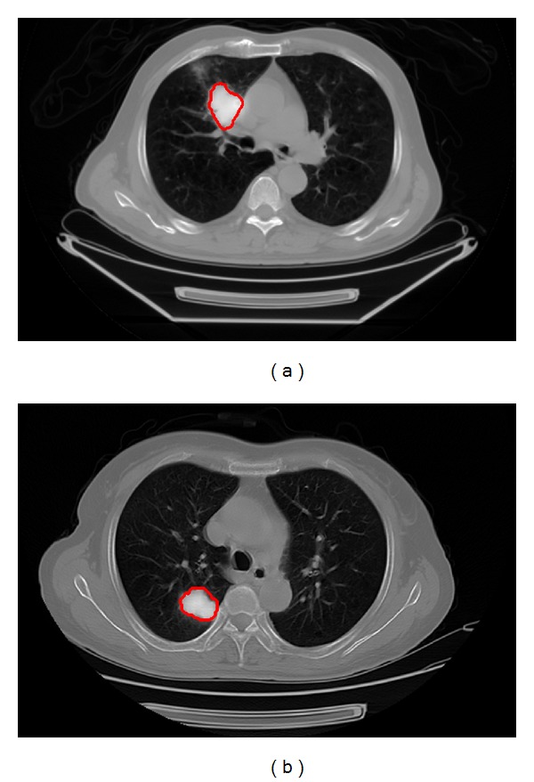
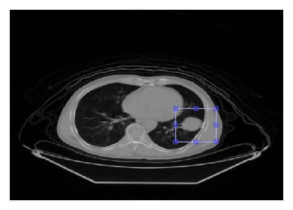
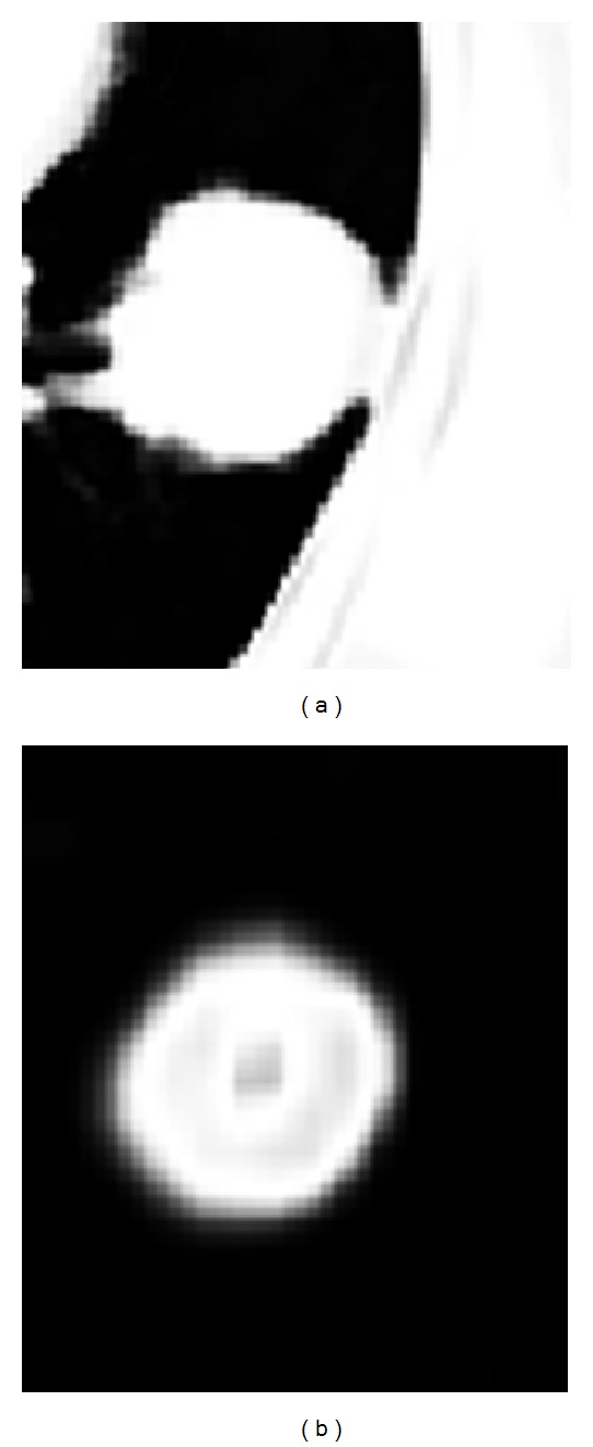
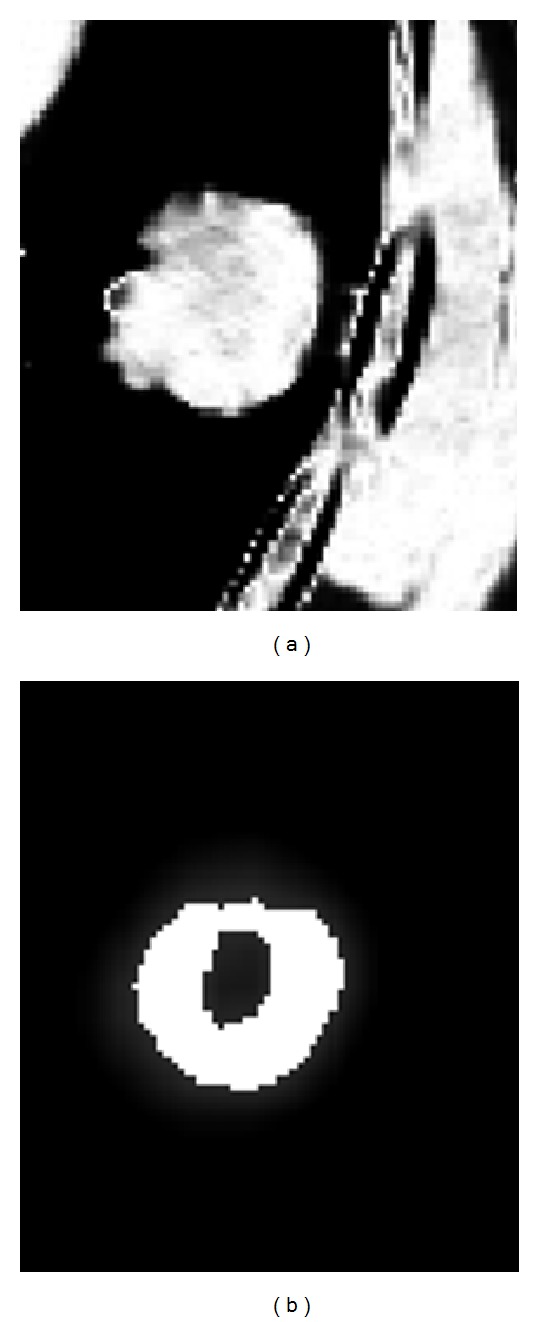
The combination of positron emission tomography (PET) and CT images provides complementary functional and anatomical information of human tissues and it has been used for better tumor volume definition of lung cancer. This paper proposed a robust method for automatic lung tumor segmentation on PET/CT images. The new method is based on fuzzy Markov random field (MRF) model. The combination of PET and CT image information is achieved by using a proper joint posterior probability distribution of observed features in the fuzzy MRF model which performs better than the commonly used Gaussian joint distribution. In this study, the PET and CT simulation images of 7 non-small cell lung cancer (NSCLC) patients were used to evaluate the proposed method. Tumor segmentations with the proposed method and manual method by an experienced radiation oncologist on the fused images were performed, respectively. Segmentation results obtained with the two methods were similar and Dice's similarity coefficient (DSC) was 0.85 ± 0.013. It has been shown that effective and automatic segmentations can be achieved with this method for lung tumors which locate near other organs with similar intensities in PET and CT images, such as when the tumors extend into chest wall or mediastinum.
正电子发射断层扫描(PET)与CT图像的结合提供了人体组织互补的功能和解剖信息,并且已被用于更精确地界定肺癌的肿瘤体积。本文提出了一种在PET/CT图像上自动分割肺肿瘤的稳健方法。新方法基于模糊马尔可夫随机场(MRF)模型。通过在模糊MRF模型中使用适当的观测特征联合后验概率分布来实现PET和CT图像信息的结合,该分布比常用的高斯联合分布表现更好。在本研究中,使用7例非小细胞肺癌(NSCLC)患者的PET和CT模拟图像来评估所提出的方法。分别采用所提出的方法和由经验丰富的放射肿瘤学家在融合图像上进行的手动方法进行肿瘤分割。两种方法获得的分割结果相似,Dice相似系数(DSC)为0.85±0.013。结果表明,对于在PET和CT图像中与其他强度相似的器官相邻的肺肿瘤,如肿瘤延伸至胸壁或纵隔时,该方法能够实现有效的自动分割。